. 2009 Aug 3:6:21.
doi: 10.1186/1743-8977-6-21.

Coal dust alters beta-naphthoflavone-induced aryl hydrocarbon receptor nuclear translocation in alveolar type II cells

Affiliations

Coal dust alters beta-naphthoflavone-induced aryl hydrocarbon receptor nuclear translocation in alveolar type II cells

Mohamed M Ghanem et al. Part Fibre Toxicol. .

Abstract

Background: Many polycyclic aromatic hydrocarbons (PAHs) can cause DNA adducts and initiate carcinogenesis. Mixed exposures to coal dust (CD) and PAHs are common in occupational settings. In the CD and PAH-exposed lung, CD increases apoptosis and causes alveolar type II (AT-II) cell hyperplasia but reduces CYP1A1 induction. Inflammation, but not apoptosis, appears etiologically associated with reduced CYP1A1 induction in this mixed exposure model. Many AT-II cells in the CD-exposed lungs have no detectable CYP1A1 induction after PAH exposure. Although AT-II cells are a small subfraction of lung cells, they are believed to be a potential progenitor cell for some lung cancers. Because CYP1A1 is induced via ligand-mediated nuclear translocation of the aryl hydrocarbon receptor (AhR), we investigated the effect of CD on PAH-induced nuclear translocation of AhR in AT-II cells isolated from in vivo-exposed rats. Rats received CD or vehicle (saline) by intratracheal (IT) instillation. Three days before sacrifice, half of the rats in each group started daily intraperitoneal injections of the PAH, beta-naphthoflavone (BNF).

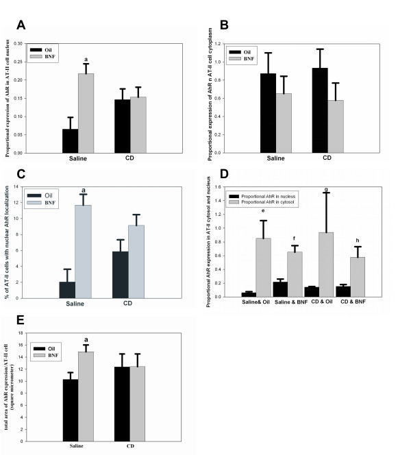
Results: Fourteen days after IT CD exposure and 1 day after the last intraperitoneal BNF injection, AhR immunofluorescence indicated that proportional AhR nuclear expression and the percentage of cells with nuclear AhR were significantly increased in rats receiving IT saline and BNF injections compared to vehicle controls. However, in CD-exposed rats, BNF did not significantly alter the nuclear localization or cytosolic expression of AhR compared to rats receiving CD and oil.

Conclusion: Our findings suggest that during particle and PAH mixed exposures, CD alters the BNF-induced nuclear translocation of AhR in AT-II cells. This provides an explanation for the modification of CYP1A1 induction in these cells. Thus, this study suggests that mechanisms for reduced PAH-induced CYP1A1 activity in the CD exposed lung include not only the effects of inflammation on the lung as a whole, but also reduced PAH-associated nuclear translocation of AhR in an expanded population of AT-II cells.


Figures

Figure 1
Figure 1
Identification of isolated AT-II cells with phosphine 3 R fluorescent dye. (A) A representative image from CD-exposed rat showing the staining of AT-II cell with bright green color. Note the presence of a hypertrophied AT-II cell (white arrow) that is larger in size than the other cells (blue arrows). Cells that are not AT-II cells (yellow arrow) do not get the stain. Reference bar is 20 μm. (B) An image of an AT-II cell captured under higher magnification demonstrating the lamellar bodies and the cells close to it do not have lamellar bodies and were classified as non AT-II cells. Reference bar is 20 μm. (C) A representative electron microscope image of an AT-II cell showing the lamellar bodies (black arrow heads). Reference bar is 2 μm.
Figure 2
Figure 2
The number (A) and purity (B) of AT-II cells in different treatment groups are shown. No significant changes in the number or purity of isolated type II cells among groups were observed. Results are means ± SE, n = 4 in all groups except in saline plus oil group (n = 3).
Figure 3
Figure 3
Representative immunofluorescent images showing the expression of AhR in AT-II cells in rats exposed to saline or CD with and without BNF. In A, B, C and D the AhR stained red, the nuclear area stained green, and the overlapping of the green area by red area produces yellow color representing localization of AhR in the nucleus. (A) An image of AhR and Cytox green from saline plus oil group. (B) An image of AhR and Cytox green from saline plus BNF group showing distinct localization of AhR in AT-II cell nucleus (yellow color). (C) An image of AhR and Cytox green from CD plus oil group. (D) An image of AhR and Cytox green from CD plus BNF group showing localization of AhR in AT-II cell nucleus (yellow color). Reference bar is 20 μm.
Figure 4
Figure 4
Morphometric quantification of AhR in the nucleus and cytoplasm of isolated AT-II cells. (A) The proportional AhR in the nucleus was significantly increased (letter a; p < 0.05) in the saline plus BNF group compared to the saline plus oil group but no increase in nuclear AhR was observed after BNF in co-treated rats. (B) No significant change in the proportional AhR expression in cytoplasm of rats receiving BNF compared to those receiving oil was observed in either the IT saline or CD groups. (C) The percentage of AT-II cells with AhR localized in the nucleus was significantly increased (letter a; p < 0.05) in BNF-exposed rats without CD exposure but not in BNF and CD exposed rats. (D) The proportional AhR expression in AT-II cell cytosol was significantly higher (P < 0.05) than that in the nucleus in all treatment groups (letters e, f, g, and h, respectively). (E) The total area of AhR expression per AT-II cell was significantly increased (p < 0.05) in rats receiving saline plus BNF compared to those receiving saline plus oil (letter a; p < 0.05) but BNF causes no changes in rats receiving CD. Results are mean + SE, n = 4 in all groups except in saline plus oil group (n = 3).

Similar articles

References

    1. Castranova V. From coal mine dust to quartz: mechanisms of pulmonary pathogenicity. Inhal Toxicol. 2000;12:7–14. doi: 10.1080/08958370050164842. - DOI - PubMed
    1. Alexandrov K, Cascorbi I, Rojas M, Bouvier G, Kriek E, Bartsch H. CYP1A1 and GSTM1 genotypes affect benzo[a]pyrene DNA adducts in smokers' lung: comparison with aromatic/hydrophobic adduct formation. Carcinogenesis. 2002;23:1969–1977. doi: 10.1093/carcin/23.12.1969. - DOI - PubMed
    1. Ghanem MM, Porter D, Battelli LA, Vallyathan V, Kashon ML, Ma JY, Barger MW, Nath J, Castranova V, Hubbs AF. Respirable coal dust particles modify cytochrome P4501A1 (CYP1A1) expression in rat alveolar cells. Am J Respir Cell Mol Biol. 2004;31:171–183. doi: 10.1165/rcmb.2003-0425OC. - DOI - PubMed
    1. Ghanem MM, Battelli LA, Mercer RR, Scabilloni JF, Kashon ML, Ma JY, Nath J, Hubbs AF. Apoptosis and Bax expression are increased by coal dust in the polycyclic aromatic hydrocarbon-exposed lung. Environ Health Perspect. 2006;114:1367–1373. - PMC - PubMed
    1. Kasper M, Rudolf T, Verhofstad AA, Schuh D, Muller M. Heterogeneity in the immunolocalization of cytokeratin-specific monoclonal antibodies in the rat lung: evaluation of three different alveolar epithelial cell types. Histochemistry. 1993;100:65–71. doi: 10.1007/BF00268879. - DOI - PubMed

LinkOut - more resources