. 2009 Aug 5;97(3):825-34.
doi: 10.1016/j.bpj.2009.05.037.

Modulation of titin-based stiffness by disulfide bonding in the cardiac titin N2-B unique sequence

Affiliations

Modulation of titin-based stiffness by disulfide bonding in the cardiac titin N2-B unique sequence

Anika Grützner et al. Biophys J. .

Abstract

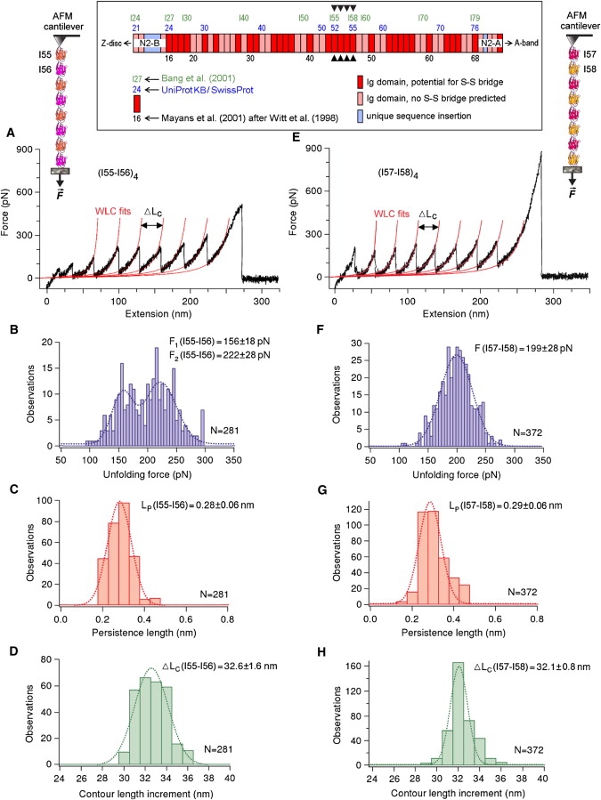
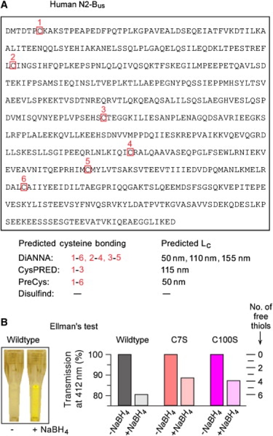
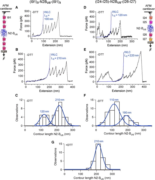
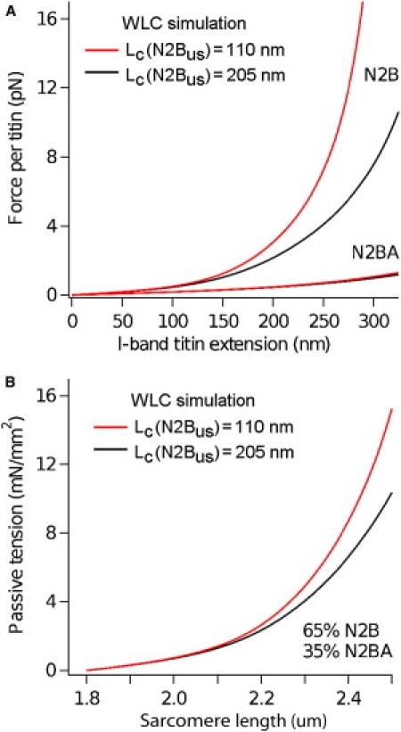
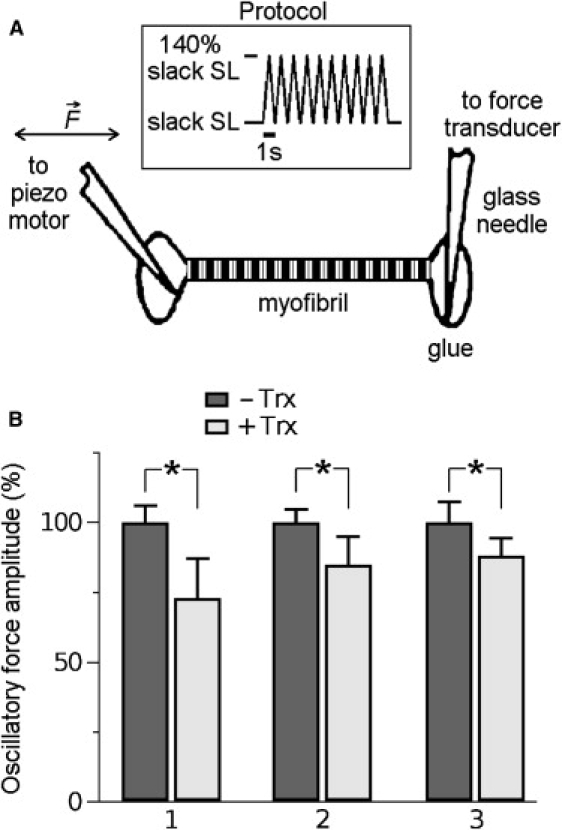
The giant protein titin is responsible for the elasticity of nonactivated muscle sarcomeres. Titin-based passive stiffness in myocardium is modulated by titin-isoform switching and protein-kinase (PK)A- or PKG-dependent titin phosphorylation. Additional modulatory effects on titin stiffness may arise from disulfide bonding under oxidant stress, as many immunoglobulin-like (Ig-)domains in titin's spring region have a potential for S-S formation. Using single-molecule atomic force microscopy (AFM) force-extension measurements on recombinant Ig-domain polyprotein constructs, we show that titin Ig-modules contain no stabilizing disulfide bridge, contrary to previous belief. However, we demonstrate that the human N2-B-unique sequence (N2-B(us)), a cardiac-specific, physiologically extensible titin segment comprising 572 amino-acid residues, contains up to three disulfide bridges under oxidizing conditions. AFM force spectroscopy on recombinant N2-B(us) molecules demonstrated a much shorter contour length in the absence of a reducing agent than in its presence, consistent with intramolecular S-S bonding. In stretch experiments on isolated human heart myofibrils, the reducing agent thioredoxin lowered titin-based stiffness to a degree that could be explained (using entropic elasticity theory) by altered extensibility solely of the N2-B(us). We conclude that increased oxidant stress can elevate titin-based stiffness of cardiomyocytes, which may contribute to the global myocardial stiffening frequently seen in the aging or failing heart.


Figures

Figure 1
Figure 1
AFM force-extension experiments to test whether titin Ig domains can be stabilized by internal (S-S) bonding. (A and E) Exemplary recordings and WLC fits to Ig-unfolding peaks; (B and F) Ig-domain unfolding forces; (C and G) persistence length, Lp, of the (partially) unfolded polypeptide chain; and (D and H) contour-length increment upon Ig unfolding, ΔLc, for two different polyprotein constructs: (AD) (I55-I56)4 and (EH) (I57-I58)4. Values at the top of the histograms are mean ± SD; dotted lines are the best Gaussian fits. (Inset) Domain numbering for the differentially spliced I-band Ig-domain segment in human cardiac titin is according to Bang et al. (23), Mayans et al. (18) using the nomenclature of Witt et al. (20), or the UniProtKB/Swiss-Prot entry (Q8WZ42) for human titin. Predictions for S-S bridge potential in Igs were taken from Mayans et al. (18). Arrowheads point to the four Ig domains investigated by AFM.
Figure 2
Figure 2
S-S bonding predictions for the human N2-Bus and Ellman's test for the presence of free thiols in recombinant N2-Bus constructs. (A) Amino-acid sequence of the human N2-Bus (accession No. X90568 in GenBankTM/EBI Data Bank), with cysteines boxed and numbered. The bottom part lists the results of four different web-based disulfide bridge-prediction algorithms calculating the probability of S-S bonding in the N2-Bus. Shown are predicted connectivities (cysteine numbers in red) and the respective contour length of N2-Bus, Lc, for each bonding prediction, assuming 0.36 nm per amino acid. (B) Results of Ellman's test to detect free thiols in recombinant wild-type and mutant (C7S; C100S) human N2-Bus constructs in solution. (Left) Example of a color change of wild-type N2-Bus in Ellman's reagent, in the presence versus absence of reducing agent, NaBH4. (Right) Relative decrease in transmission at 412 nm measured for wild-type and mutant N2-Bus constructs under reducing and nonreducing conditions; a decrease in transmission indicates that thiols became free in the presence of NaBH4 (scale on right).
Figure 3
Figure 3
AFM force-extension experiments probing the presence of S-S bonds in the N2-Bus. Polyproteins (I91)3-N2-Bus-(I91)3 (left panels) and (I24)-(I25)-N2-Bus-(I26)-(I27) (right panels) were recombinantly expressed and stretched by AFM. Only those recordings that showed at least four regularly spaced Ig-unfolding peaks for (I91)3-N2-Bus-(I91)3 or three regularly spaced Ig-unfolding peaks for (I24)-(I25)-N2-Bus-(I26)-(I27) were analyzed by WLC fitting, because only then could we be confident that the whole N2-Bus had been stretched. (AE) Exemplary recordings in 200 mM PBS buffer lacking reducing agent (−DTT), and WLC fit of N2-Bus force-extension behavior up to the first Ig-unfolding peak. Calculated contour length (Lc) values for the N2-Bus are indicated in blue text. (C and F) Histograms showing bimodal contour-length distribution in the absence of reducing agent (−DTT). Blue lines and values are single Gaussian fits and mean Lc. (G) Histogram of contour-length distribution for the N2-Bus in 200 mM PBS buffer supplemented with 10 mM DTT (+DTT). Blue line and value indicate single Gaussian fit and mean Lc. Note the absence of shorter contour lengths.
Figure 4
Figure 4
WLC simulations of the effect of a less-extensible N2-Bus on titin-based spring force. (A) Predicted differences in the force/titin versus I-band titin extension curves using Lc values for the N2-Bus of either 110 nm (red curves) or 205 nm (black curves). The simulation was performed separately for the two human cardiac titin isoforms, N2B (3000 kDa) and N2BA (3300 kDa). (B) Predicted difference in the passive tension versus SL relationship of human cardiac titin, using a contour length for the N2-Bus of either 110 nm (red curve) or 205 nm (black curve). This simulation also considered the 35:65 expression ratio of N2BA:N2B titin isoforms in normal human left ventricle.
Figure 5
Figure 5
Effect of reducing agent on the passive stiffness of cardiomyofibrils isolated from human donor hearts. (A) Experimental setup and mechanics protocol. (B) Change in the passive stiffness of three different myofibrils studied in relaxing buffer supplemented with 50 nM Trx reductase and 2 mM NADPH, before (−Trx) and after (+Trx) application of human Trx (8 μM). Bars show average oscillatory force amplitudes (mean ± SE) in bursts of 10 stretch-release cycles; data were expressed relative to the mean amplitude before addition of Trx. Time between (−Trx) and (+Trx) recordings is ∼5 min. Asterisks indicate statistically significant differences (p < 0.05 in Student's t-test).

References

    1. Granzier H., Labeit S. Structure-function relations of the giant elastic protein titin in striated and smooth muscle cells. Muscle Nerve. 2007;36:740–755. - PubMed
    1. Linke W.A. Sense and stretchability: the role of titin and titin-associated proteins in myocardial stress-sensing and mechanical dysfunction. Cardiovasc. Res. 2008;77:637–648. - PubMed
    1. Neagoe C., Kulke M., del Monte F., Gwathmey J.K., de Tombe P.P. Titin isoform switch in ischemic human heart disease. Circulation. 2002;106:1333–1341. - PubMed
    1. Nagueh S.F., Shah G., Wu Y., Torre-Amione G., King N.M. Altered titin expression, myocardial stiffness, and left ventricular function in patients with dilated cardiomyopathy. Circulation. 2004;110:155–162. - PubMed
    1. Makarenko I., Opitz C.A., Leake M.C., Neagoe C., Kulke M. Passive stiffness changes caused by upregulation of compliant titin isoforms in human DCM hearts. Circ. Res. 2004;95:708–716. - PubMed

Publication types

MeSH terms

LinkOut - more resources