Review
. 2009 Aug;19(8):364-74.
doi: 10.1016/j.tcb.2009.05.007. Epub 2009 Aug 3.

Scaffolds: interaction platforms for cellular signalling circuits

Affiliations
Review

Scaffolds: interaction platforms for cellular signalling circuits

András Zeke et al. Trends Cell Biol. 2009 Aug.

Abstract

Scaffold proteins influence cellular signalling by binding to multiple signalling enzymes, receptors or ion channels. Although normally devoid of catalytic activity, they have a big impact on controlling the flow of signalling information. By assembling signalling proteins into complexes, they play the part of signal processing hubs. As we learn more about the way signalling components are linked into natural signalling circuits, researchers are becoming interested in building non-natural signalling pathways to test our knowledge and/or to intentionally reprogram cellular behaviour. In this review, we discuss the role of scaffold proteins as efficient tools for assembling intracellular signalling complexes, both natural and artificial.


Figures

Figure 1
Figure 1
Ste5, a classical signalling scaffold. (a) Our current view on the scaffolding interactions of the yeast Ste5 protein. Ste4 and Ste18 are the β and γ subunits of the heterotrimeric G-protein with a role in recruiting components of the pathway to the plasma membrane upon pheromone receptor activation. Ste20, Ste11, Ste7 and Fus3 are protein kinases, phosphorylating each other in successive steps (illustrated with green arrows). The Fus3 MAP kinase, as the output of the scaffolded module, phosphorylates substrate targets (such as the cyclin-dependent kinase inhibitor Far1). Other proteins, such as Ste50 and Bem1, have additional regulatory roles in this cascade. (b) The Ste5 signalling scaffold in its PPI network context. Proteins possessing intrinsic enzymatic activity are coloured orange, passive components are gray and the edges of the full 3-graphs that represent scaffolded signalling complexes are drawn with a thick line. These interactions were taken directly from the STRING database (http://string.embl.de), with the following settings: confidence level = 0.90, type: experimental only, maximum interactors = 100. (Some interacting partners are not presented for simplicity.).
Figure 2
Figure 2
Schematic representation of the four main scaffold mechanisms. Known examples of signalling scaffold proteins display four distinct mechanisms through which they can modify signalling between active components. (a) Scaffolds tether enzymes close in space and enhance effective local concentrations. (b) They can mediate assembly of signalling complexes in a combinatorial manner, meaning that a certain active component (depicted as red triangle) can participate in signalling through different pathways using distinct scaffold proteins. (c) The function of full signalling modules can be dynamically regulated if the turnover or accessibility of the scaffold is dynamically regulated, without the need to execute the same type of regulation individually on the active components. (d) Some scaffolds can also modify the conformation of enzymes binding to them, or in turn the conformation of the scaffold can also be modified.
Figure 3
Figure 3
Scaffold proteins regulate characteristics of intracellular signalling in space, time or input strength. Top panels (a–c) display presumed signalling profiles of certain circuits in the absence (blue) or in the presence (red) of a scaffold. In (d), signalling scenarios are compared before and after scaffold modification took place through feedback. Panels in the middle rows schematically depict the circuits that generate behaviours shown in the top panels. Bottom panels demonstrate examples. (a) Scaffolds can provide highly localized signalling. AKAP proteins confine the activity of protein kinase A (PKA) into well-defined cellular regions, and they also help inactivate the adenyl cyclase-generated cAMP signal through binding to distinct phosphodiesterase (PDE) isoforms [9]. The local assembly of all these signalling elements serves to create localized ‘pulses’ of cAMP-dependent phosphorylations that translates into perinuclear Ca2+-pulses in the case of mAKAP [10]. (b) Scaffolds can control the dynamics of signalling. In the phototransduction system of the fly Drosophila melanogaster, the seven-transmembrane-type rhodopsin photoreceptor and its downstream effectors (including the TRP ion channel) are organized into a single multimolecular complex with the help of the multi-PDZ protein InaD (Inactivation no afterpotential D). When the receptor is switched on by light, it acts as an exchanger of GDP for GTP on its associated heterotrimeric G-protein, dissociating it into α- and β–γ subunits. The binding of free β–γ subunit activates phospholipase C (PLC), which in turn hydrolyses the phosphoinositides contained in the membrane to inositol (1,4,5)-trisphosphate [Ins(1,4,5)P3] and diacylglycerol (DAG). Although Ins(1,4,5)P3 seems to have no direct role in this system, DAG activates both the transient receptor potential (TRP) channels to generate Ca2+-influx and the enzyme protein kinase C (PKC). Once turned on, the latter readily phosphorylates the TRP channel, rendering it inactive, thus terminating the signal. The recruitment of both positive effectors (phospholipase C) and feed-forward inhibitors (PKC) into a signalling complex accounts for the generation of ion flux pulses separated in time by barely a few milliseconds – creating one of the fastest known heterotrimeric G-protein-based signalling cascades [11]. (c) Scaffolds can change dose–response relationships. The mating pathway of S. cerevisiae relies on the presence of the scaffold protein Ste5, which organizes the protein kinases Ste11, Ste7 and the Fus3 MAP kinase into a single complex. Surprisingly, after binding to Ste5, Fus3 becomes a 5000-fold better substrate for its upstreamkinase, Ste7. It seems that Ste5makes Fus3 a better substrate by unlocking its activation loop for more efficient phosphorylation by Ste7 [13]. (d) Scaffolds can provide memory effects. In addition to the immediate feed-forward mechanisms in Drosophila photoreception, the presence of the scaffold enables a sophisticated mechanism for the adaptation to high-input and low-input conditions. In low-light conditions, the fifth PDZ domain of the InaD protein resides in an open conformation. During repeated stimulation of the pathway (under high-light conditions), the activation of PKC also results in the phosphorylation of the scaffold, which in turn suffers a conformational change, turning the fifth PDZ domain into a closed conformation, and releasing its previously bound partner – most probably PLC. This decreases the flux through the pathway, resulting in long-term adaptation [12].
Figure 4
Figure 4
Scaffold-based network architectures with diverse pathway dynamics. Top panels show experimental kinetic profiles for circuits with wild-type Ste5 (blue) and with a synthetic Ste5 scaffold (red). Lower panels schematically depict underlying circuit architectures. The Ste5 scaffold protein was engineered to include an additional binding site (e.g. by addition of a leuzine zipper, shown in magenta) to enable it to bind other proteins containing a complementary protein interaction element (e.g. another leuzine zipper). By recruiting various effectors or decoys to this scaffolded MAP-kinase module it was possible to change yeast mating pathway kinetics at will [43]. (The original components of this MAPK module: Ste11, Ste7 and Fus3 kinases are coloured in different shades of orange.) (a) Expression of a positive activator (shown in light blue) through a promoter that is responsive to pathway flux creates a simple positive feedback loop, providing more rapid responses compared with wild-type cells. (b) If pathway flux triggers the expression of a negative regulator (yellow), then a simple negative-feedback loop will result. This creates a pulse-like response upon stimulation, unlike wild-type yeast that exhibits a continuous, saturated response. (c) With a careful combination of positive- and negative-feedback elements almost any desired dynamic profile can be realized. A good example is the combination of a constitutively expressed positive regulatory element with a signal-inducible negative element. The two proteins naturally compete with each other for the same binding site, and the inhibitor element will gradually displace the enhancer element as the signalling pathway becomes activated by sustained stimulus. As expected, the system provides a pulse-like output in time, but the peak is shifted towards earlier time points when compared with a simple negative-feedback loop (containing only the inducible inhibitor). Thus, the system behaves as an accelerator. (d) A more complex way to change signalling characteristics can be achieved through the use of decoys – proteins only consisting of binding sites – that, for example, can disrupt association of a constitutively expressed negative regulatory factor with the scaffold. When compared with wild-type cells, a pronounced delay of signal propagation will become apparent. The decoy gradually diminishes the inhibition of signalling as the pathway becomes activated by a sustained stimulus.
Figure I
Figure I
Enrichment of signalling molecules around a scaffold.
Figures I
Figures I
General mechanisms for the evolution of scaffolds.
Figures I
Figures I
A synthetic biology ‘toolkit’.

Comment in

  • Finding scaffold proteins in interactomes.
    Ramírez F, Albrecht M. Ramírez F, et al. Trends Cell Biol. 2010 Jan;20(1):2-4. doi: 10.1016/j.tcb.2009.11.003. Epub 2009 Dec 11. Trends Cell Biol. 2010. PMID: 20005715 No abstract available.

References

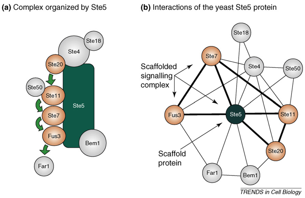
    1. Choi KY, et al. Ste5 tethers multiple protein kinases in the MAP kinase cascade required for mating in S. cerevisiae. Cell. 1994;78:499–512. - PubMed
    1. Friedman A, Perrimon N. Genetic screening for signal transduction in the era of network biology. Cell. 2007;128:225–231. - PubMed
    1. Bardwell L. A walk-through of the yeast mating pheromone response pathway. Peptides. 2005;26:339–350. - PMC - PubMed
    1. Ferrrell JE, Jr, Cimprich KA. Enforced proximity in the function of a famous scaffold. Mol. Cell. 2003;11:289–291. - PubMed
    1. Bhattacharyya RP, et al. Domains, motifs, and scaffolds: the role of modular interactions in the evolution and wiring of cell signalling circuits. Annu. Rev. Biochem. 2006;75:655–680. - PubMed

Publication types