. 2009 Aug 11;106(32):13329-34.
doi: 10.1073/pnas.0901966106. Epub 2009 Jul 27.

Structural basis for a human glycosylation disorder caused by mutation of the COG4 gene

Affiliations

Structural basis for a human glycosylation disorder caused by mutation of the COG4 gene

Brian C Richardson et al. Proc Natl Acad Sci U S A. .

Abstract

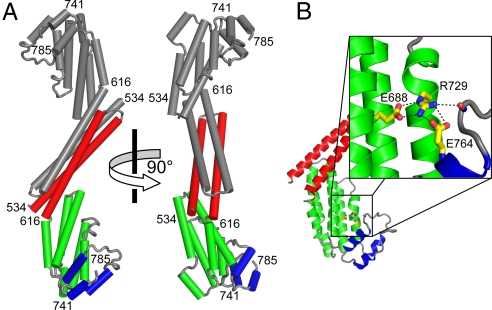
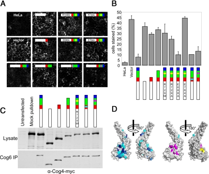
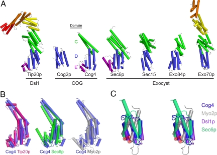
The proper glycosylation of proteins trafficking through the Golgi apparatus depends upon the conserved oligomeric Golgi (COG) complex. Defects in COG can cause fatal congenital disorders of glycosylation (CDGs) in humans. The recent discovery of a form of CDG, caused in part by a COG4 missense mutation changing Arg 729 to Trp, prompted us to determine the 1.9 A crystal structure of a Cog4 C-terminal fragment. Arg 729 is found to occupy a key position at the center of a salt bridge network, thereby stabilizing Cog4's small C-terminal domain. Studies in HeLa cells reveal that this C-terminal domain, while not needed for the incorporation of Cog4 into COG complexes, is essential for the proper glycosylation of cell surface proteins. We also find that Cog4 bears a strong structural resemblance to exocyst and Dsl1p complex subunits. These complexes and others have been proposed to function by mediating the initial tethering between transport vesicles and their membrane targets; the emerging structural similarities provide strong evidence of a common evolutionary origin and may reflect shared mechanisms of action.


Conflict of interest statement

The authors declare no conflict of interest.

Figures

Fig. 1.
Fig. 1.
X-ray crystal structure of Cog4-(525–785). (A) H. sapiens Cog4, including residues 536–785. (B) Ionic interaction network centered around Arg 729.
Fig. 2.
Fig. 2.
Mutational analysis of Cog4 function in Cog4-silenced HeLa cells. (A) Cells transfected with the indicated siRNA-resistant allele were incubated with GNL-Alexa-647, which labels mis-glycosylated cell surface proteins. All constructs (except “vector”) include Cog4 residues 1–524, indicated by the white bars. Red, green, and blue bars represent the protein regions color-coded as in Fig. 1A. Cyan and magenta boxes indicate sets of mutations as indicated in Fig. S2. A yellow box indicates mutations of R729 or interacting residues as noted on the bar itself (e.g., R729W). (B) Quantification of the percentage of cells [± SD, n = 3 (>100 cells each)] as shown in Fig. 2A with detectable GNL staining of plasma membrane glycoproteins. (C) To assess protein expression level, cells transfected with silencing-resistant Cog4 alleles carrying C-terminal 3× Myc tags were analyzed by western blotting. To assess the integrity of the COG complex, the quantity of tagged Cog4 present in Cog6 immunoprecipitation reactions was evaluated by western blotting. (D) Surface representations of the Cog4-(525–785) monomer. In the left-hand pair of images, the surface is color-coded by sequence conservation (darker blue is more conserved). In the right-hand pair, the residues that were studied by site-directed mutagenesis are color-coded to match the triangles in Fig. S2.
Fig. 3.
Fig. 3.
Mutational analysis of Cog4p function in S. cerevisiae. The percentage CPY secreted was calculated by measuring intracellular and extracellular CPY by western blotting (see Fig. S3C). The vacuolar sorting mutant vps1–1 was included as a control.
Fig. 4.
Fig. 4.
Structural alignment of Cog4-(525–785) to known COG, exocyst and Dsl1p subunits. (A) Shown are S. cerevisiae Tip20p (PDB ID 3FHN, residues 4–701 out of 701) (20), Cog2p (2JQQ, residues 109–262 out of 262) (14), Sec6p (2FJI, residues 411–805 out of 805) (18), Drosophila melanogaster Sec15 (2A2F, residues 382–699 out of 766) (19), S. cerevisiae Exo84p (2D2S, residues 525–753 out of 753) (15), and S. cerevisiae Exo70p (2PFV, residues 67–623 out of 623) (–17). Pairwise alignment was performed with the program DaliLite (47) to match each of the other structures to Cog4-(525–785). The DaliLite Z scores for the alignments shown were 12.3 (Cog4-Tip20p), 3.8 (Cog4-Cog2p), 13.1 (Cog4-Sec6p), 10.1 (Cog4-Sec15), 6.2 (Cog4-Exo84p), and 8.0 (Cog4-Exo70p). (B) Cog4- (525–785) superimposed on proteins containing similar domains C, D, and E. (C) Stereoview of E domains, aligned using DaliLite and with the N terminus of each domain indicated by a red sphere. Included, in addition to the structures cited above, are the E domains of the cargo-binding domain of S. cerevisiae Myo2p (2F6H) (33) and the Dsl1p complex subunit Dsl1p (Y. Ren, P.D.J., and F.M.H., personal communication). No significant alignment was discernable for domain E of Tip20p.

References

    1. Lupashin V, Sztul E. Golgi tethering factors. Biochim Biophys Acta. 2005;1744:325–339. - PubMed
    1. Ungar D, Oka T, Krieger M, Hughson FM. Retrograde transport on the COG railway. Trends Cell Biol. 2006;16:113–120. - PubMed
    1. Zolov SN, Lupashin VV. Cog3p depletion blocks vesicle-mediated Golgi retrograde trafficking in HeLa cells. J Cell Biol. 2005;168:747–759. - PMC - PubMed
    1. Walter DM, Paul KS, Waters MG. Purification and characterization of a novel 13 S hetero-oligomeric protein complex that stimulates in vitro Golgi transport. J Biol Chem. 1998;273:29565–29576. - PubMed
    1. Chatterton JE, et al. Expression cloning of LDLB, a gene essential for normal Golgi function and assembly of the ldlCp complex. Proc Natl Acad Sci USA. 1999;96:915–920. - PMC - PubMed

Publication types

MeSH terms

Substances

Associated data