. 2009 Aug 4;106(31):13010-5.
doi: 10.1073/pnas.0903691106. Epub 2009 Jul 27.

Inclusion formation and neuronal cell death through neuron-to-neuron transmission of alpha-synuclein

Affiliations

Inclusion formation and neuronal cell death through neuron-to-neuron transmission of alpha-synuclein

Paula Desplats et al. Proc Natl Acad Sci U S A. .

Erratum in

  • Proc Natl Acad Sci U S A. 2009 Oct 13;106(41):17606

Abstract

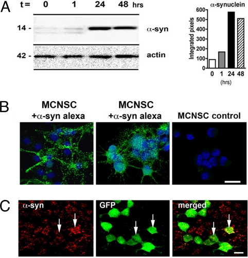
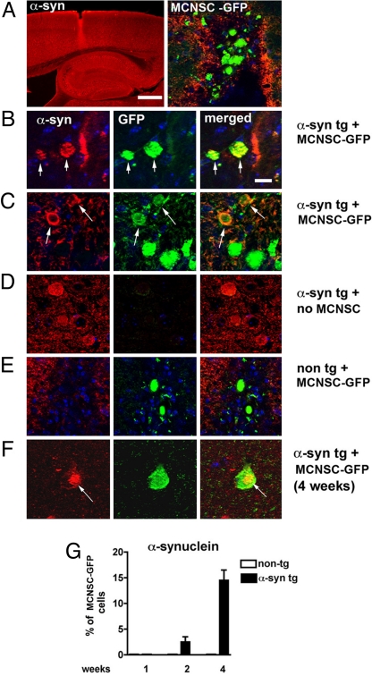
Neuronal accumulation of alpha-synuclein and Lewy body formation are characteristic to many neurodegenerative diseases, including Parkinson's disease (PD). This Lewy pathology appears to spread throughout the brain as the disease progresses. Furthermore, recent studies showed the occurrence of Lewy pathology in neurons grafted into the brains of PD patients, suggesting the spread of pathology from the host tissues to the grafts. The mechanism underlying this propagation is unknown. Here, we show that alpha-synuclein is transmitted via endocytosis to neighboring neurons and neuronal precursor cells, forming Lewy-like inclusions. Moreover, alpha-synuclein was transmitted from the affected neurons to engrafted neuronal precursor cells in a transgenic model of PD-like pathology. Failure of the protein quality control systems, especially lysosomes, promoted the accumulation of transmitted alpha-synuclein and inclusion formation. Cells exposed to neuron-derived alpha-synuclein showed signs of apoptosis, such as nuclear fragmentation and caspase 3 activation, both in vitro and in vivo. These findings demonstrate the cell-to-cell transmission of alpha-synuclein aggregates and provide critical insights into the mechanism of pathological progression in PD and other proteinopathies.


Conflict of interest statement

The authors declare no conflict of interest.

Figures

Fig. 1.
Fig. 1.
α-Synuclein uptake by mouse cortical neuronal stem cells (MCNSCs). (A) Immunoblot analysis of extracellular α-synuclein uptake after 24 and 48 h in MCNSCs. (B) Confocal microscopy analysis demonstrating the MCNSC uptake of Alexa-Fluor-488-tagged extracellular α-synuclein (green). (C) The MCNSCs infected with a lentivirus-GFP (green) were cocultured with a neuronal cell line (B103) infected with a lentivirus expressing α-synuclein (red). Punctate and diffuse red staining represents the transmitted α-synuclein in acceptor cells (arrows). (Scale bars, 10 μm.)
Fig. 2.
Fig. 2.
Transmission of α-synuclein from host to grafted neural stem cells. (A) Thy-1 α-synuclein transgenic mice received stereotaxic intrahippocampal injections of mouse cortical neuronal stem cells (MCNSCs) (150,000 cells) infected with lentivirus-GFP. Confocal microscopy analysis demonstrates the site of the injection and the GFP-tagged MCNSCs. The α-synuclein is immunolabeled with tyramide red. (B and C) Colocalization of α-synuclein (red) in acceptor MCNSCs (green) (arrows) in areas adjacent to the injection site. (Scale bar, 10 μm.) (D) Control experiment showing the background fluorescence in the absence of MCNSC injection. (E) Control experiments showing lack of human α-synuclein in MCNSCs injected into nontransgenic mice. (F) A compact inclusion body with human α-synuclein immunoreactivity (red) 4 weeks postinjection. (G) Quantification of human α-synuclein-positive MCNSCs grafted into Thy-1 α-synuclein transgenic mice or nontransgenic control mice.
Fig. 3.
Fig. 3.
Inclusion body formation via cell-to-cell transmission of α-synuclein. (A) Differentiated SH-SY5Y neuronal cells overexpressing myc-tagged α-synuclein (green) were cocultured with SH-SY5Y acceptor cells labeled with Qtracker 585 (Q; red). (Upper) Diffuse distribution of transmitted α-synuclein in acceptor cells. (Lower) Inclusion body formation (arrow) in acceptor cells. (Scale bars, 20 μm.) (Left) Percentage of acceptor cells with transmitted α-synuclein. (Right) Percentage of acceptor cells with compact inclusion body. Three independent experiments were performed, and on average, 300 cells per sample were analyzed. Error bars are SEM (*, P < 0.05; **, P < 0.01). Inclusion bodies were defined as distinct clusters of α-synuclein-positive structures near the nucleus with diameters exceeding 2 μm. (B) Time-dependent transmission of α-synuclein and inclusion body formation. The experiments were set up in the same way as in (A). The cocultures were examined at indicated times. For quantification, at least 200 cells (average 305 cells) were examined, and four independent experiments were performed. (C) Characterization of inclusion bodies in acceptor cells. (Upper) Ubiquitin-positive inclusion bodies in acceptor cells (arrows). (Scale bar, 10 μm.) (Lower) Thioflavin-S-positive inclusion bodies in acceptor cells (arrowhead, cell outlined with broken line). (Scale bar, 20 μm.) The graph represents the percentage of inclusion bodies labeled with anti-ubiquitin antibodies or thioflavin S. Three independent experiments were performed, examining 8–14 inclusions per experiment. Error bars are SEM. (D) Membrane leakage analysis. The cells overexpressing the indicated proteins were subjected to the lactate dehydrogenase (LDH) release assay. All of the infected cells show lower levels of LDH release compared with the noninfected cells.
Fig. 4.
Fig. 4.
Increased deposition of transmitted α-synuclein by lysosomal failure. Differentiated SH-SY5Y cells were incubated with conditioned medium containing secreted myc-α-synuclein for 2 days. Lysosomal (Baf A1) and proteasomal (MG132) inhibitors were added at the final 16 h of treatment. The cells were immunostained with anti-myc antibody. (A) Fluorescence intensities measured from individual cells. (B) Number of cells with inclusion bodies. Three experiments were performed, and 100 cells were analyzed per sample. Error bars are SEM (*, P < 0.05; **, P < 0.01).
Fig. 5.
Fig. 5.
Degeneration of primary neurons and mouse cortical neuronal stem cells (MCNSCs) after exposure to the neuronal-cell-derived extracellular α-synuclein. (A) Nuclear fragmentation in the primary cortical neurons after incubation with α-synuclein medium and LacZ medium. Four independent experiments were performed, and on average, 190 cells were analyzed per sample. Error bars are SEM (*, P < 0.05; **, P < 0.01). (B) Quantitative immunofluorescence of activated caspase 3 in the primary cortical neurons after the same incubation as in (A). (C) Caspase 3 activation in the MCNSCs grafted into the Thy-1 α-synuclein transgenic mice. The proportions of the GFP-positive cells that exhibited activated caspase 3 were analyzed at the different time points.

Comment in

  • Is Parkinson's disease a prion disorder?
    Olanow CW, Prusiner SB. Olanow CW, et al. Proc Natl Acad Sci U S A. 2009 Aug 4;106(31):12571-2. doi: 10.1073/pnas.0906759106. Epub 2009 Jul 28. Proc Natl Acad Sci U S A. 2009. PMID: 19666621 Free PMC article. No abstract available.

References

    1. Kim C, Lee S-J. Controlling the mass action of α-synuclein in Parkinson's disease. J Neurochem. 2008;107:303–316. - PubMed
    1. Lesage S, Brice A. Parkinson's disease: From monogenic forms to genetic susceptibility factors. Hum Mol Genet. 2009;18:R48–R59. - PubMed
    1. Spillantini MG, Crowther RA, Jakes R, Hasegawa M, Goedert M. α-Synuclein in filamentous inclusions of Lewy bodies from Parkinson's disease and dementia with Lewy bodies. Proc Natl Acad Sci USA. 1998;95:6469–6473. - PMC - PubMed
    1. Maries E, Dass B, Collier TJ, Kordower JH, Steece-Collier K. The role of α-synuclein in Parkinson's disease: Insights from animal models. Nat Rev Neurosci. 2003;4:727–738. - PubMed
    1. Cookson MR. The biochemistry of Parkinson's disease. Annu Rev Biochem. 2005;74:29–52. - PubMed

Publication types

Substances