. 2009 Aug 11;106(32):13164-8.
doi: 10.1073/pnas.0906429106. Epub 2009 Jul 27.

DNA-mediated redox signaling for transcriptional activation of SoxR

Affiliations

DNA-mediated redox signaling for transcriptional activation of SoxR

Paul E Lee et al. Proc Natl Acad Sci U S A. .

Abstract

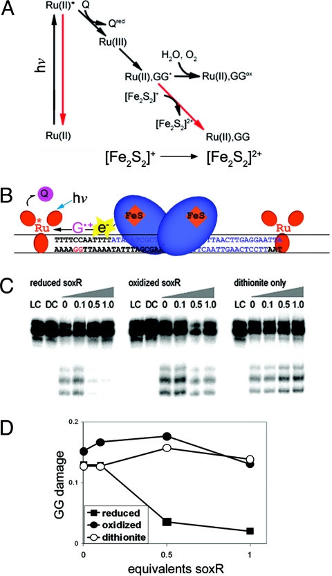
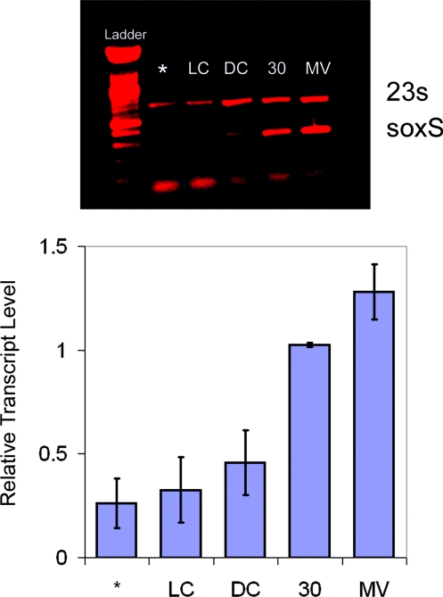
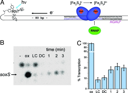
In enteric bacteria, the cellular response to oxidative stress is activated by oxidation of the iron-sulfur clusters in SoxR, which then induces transcription of soxS, turning on a battery of defense genes. Here we demonstrate both in vitro and in cells that activation of SoxR can occur in a DNA-mediated reaction with guanine radicals, an early genomic signal of oxidative stress, serving as the oxidant. SoxR in its reduced form is found to inhibit guanine damage by repairing guanine radicals. Moreover, cells treated with a DNA-binding photooxidant, which generates guanine radicals, promotes the expression of soxS. In vitro, this photooxidant, tethered to DNA 80 bp from the soxS promoter, induces transcription by activating SoxR upon irradiation. Thus, transcription can be activated from a distance through DNA-mediated charge transport. This chemistry offers a general strategy for DNA-mediated signaling of oxidative stress.


Conflict of interest statement

The authors declare no conflict of interest.

Figures

Fig. 1.
Fig. 1.
Schematized model of transcriptional activation of SoxR from a distance through DNA-mediated charge transport. Here a tethered metal complex (yellow) is used to inject an electron hole into the DNA base pair stack (dark blue) so as to generate a guanine radical (red) (1). DNA-mediated charge transport from SoxR (light blue) (2), bound at its promoter site, to the guanine radical fills the hole and leads to oxidation and activation of SoxR. The SoxR structure shown is based upon the crystal structure of oxidized SoxR bound to DNA (10).
Fig. 2.
Fig. 2.
Inhibition of guanine damage by reduced SoxR. (A) In the Ru flash quench scheme, ground state Ru(II) bound to DNA is irradiated at 442 nm to form excited Ru(II)*. The excited state is quenched by [Co(NH3)5Cl]2+ (Q), resulting in Ru(III) in situ, a ground state oxidant able to abstract an electron from guanine doublets in DNA. If the resulting guanine radical is allowed to react with water, permanent damage products, such as 8-oxo-guanine, form (black arrows). Pathways that prevent formation of damage (red arrows) are recombination processes and/or donation of an electron from the reduced [2Fe2S]+ cluster of SoxR to form [2Fe2S]2+. (B) The experimental system is shown schematically (see Methods). SoxR (blue) is bound to its promoter site on DNA. [Ru(phen)(dppz)(bpy′)]2+ (red), noncovalently intercalated into DNA, is able to oxidize a guanine residue, and the [2Fe2S]+ cluster of SoxR can donate an electron to fill the resulting hole. (C) Denaturing PAGE analysis of guanine oxidation is shown. The DNA used is a 48-mer containing the SoxR binding site. The DNA is 32P-labeled on the 5′-end of the GG containing strand. Concentrations used are 0.5 μM DNA, 5 μM [Ru(phen)(dppz)(bpy′)]2+, and 500 μM [Co(NH3)5Cl]2+. To reduce SoxR, dithionite is added at 10-fold the concentration of SoxR. LC indicates the absence of Ru; DC indicates the absence of light; numbers indicate equivalents of E. coli SoxR:DNA or, for dithionite only, the amount of reductant that would accompany added SoxR. Samples were irradiated for 20 min at 442 nm using a He/Cd laser. After reaction, DNA samples were treated with piperidine to reveal DNA damage as strand breaks; the band above the parent band reflects incomplete reaction with piperidine. The lower bands reflect characteristic oxidative damage at the 5′-GGA-3′ sites. While some decreased damage is evident at very high protein ratios, both with oxidized and reduced protein, consistent with competition between the protein and non-covalent ruthenium for DNA, significant inhibition of damage is evident preferentially for reduced SoxR. D. Quantitation of damage at guanines is shown standardized against the parent band. Uncertainties in the measurement are estimated to be 5%. Guanine damage is greatly attenuated as increasing amounts of reduced SoxR are added to the DNA. This effect is not seen with either oxidized SoxR or dithionite only.
Fig. 3.
Fig. 3.
SoxS expression in E.coli induced by treatment with [Rh(phi)2bpy]3+, a DNA photooxidant, or with methyl viologen. (Upper) An agarose gel image of RT-PCR products stained with ethidium is shown. The top band is a 23S internal control. The bottom band is the soxS transcript. The lowest molecular weight band is free primer. Cells were grown in the presence of 50 μM [Rh(phi)2bpy]Cl3or 50 μM methyl viologen. The asterisk (*) indicates cells that were grown without [Rh(phi)2bpy]Cl3 and not irradiated. LC and DC indicate, respectively, samples irradiated in the absence of metal or treated with metal but not irradiated. The lane labeled 30 represents a culture grown in the presence of [Rh(phi)2bpy]3+ and irradiated for 30 min in 24-well plates without shaking. MV indicates a sample treated with 50 μM methyl viologen and shaken continuously for 30 min at 250 rpm. All irradiations were carried out using a solar simulator with a UV filter and a 1-kW Hg/Xe lamp. LC samples were irradiated for 30 min. (Lower) Levels of soxS bands quantified as the ratio of soxS transcript to the 23S internal control. The 1-tailed P values for the 30 min and MV samples are less than 0.001 compared to the LC. Error bars represent the standard error of the average of 4 separate trials.
Fig. 4.
Fig. 4.
Transcriptional activation of SoxR from a distance triggered by photoactivation of [Rh(phi)2bpy′]3+ tethered to a 180-mer DNA duplex containing the soxS promoter region. (A) Schematic of the SoxR/DNA complex used to carry out the abortive transcription assay. The SoxR binding site is shown in blue. All experiments were carried out anaerobically. Photooxidation of DNA by the covalently tethered [Rh(phi)2bpy′]3+ oxidizes reduced SoxR bound to DNA. The samples were then incubated with RNA polymerase, and transcription was initiated by addition of a buffer containing a starting dinucleotide, [ApG], ATP, and radiolabeled UTP. The resulting mRNA product is the 4-mer RNA radiolabeled as indicated by the asterisk. Reinitiation of transcription is inhibited by the presence of heparin in the buffer. DNA (30 nM) and 300 nM SoxR are used. Both E. coli and P. aeruginosa SoxR were used (see Methods). P. aeruginosa SoxR was found to show greater stability. (B) Denaturing PAGE of mRNA products of transcriptional activation by SoxR. Shown are the products formed in the absence of SoxR, “-”; in the presence of aerated, oxidized SoxR, “ox”; the DNA template lacking the covalently tethered [Rh(phi)2bpy′]3+ but with reduced SoxR and irradiated, “LC”; the sample with reduced SoxR and covalent Rh/DNA but without irradiation, “DC”; or with reduced SoxR and covalent Rh/DNA and irradiated for increasing time in minutes, 1, 2, 3; samples treated for longer times gave variable precipitation. The top band reflects background transcription and is inhibited by DNA-bound SoxR; the bottom band is the soxS 4-mer transcript. Samples were irradiated in an anaerobic glovebox with a fiber optic cable attached to an LCD lamp (350 nm) (see Methods). (C) Quantitation of soxS expression is given as the percent transcription found versus that with fully oxidized SoxR; data were normalized to the background activity in the dark control (DC). The values for the irradiated samples have 1-tailed P values versus the light control of 0.106, 0.060, and 0.052 for 1, 2, and 3 min, respectively. The error bars represent the standard error of the average of 4 separate trials.

References

    1. Imlay JA. Cellular defenses against superoxide and hydrogen peroxide. Annu Rev Biochem. 2008;77:755–776. - PMC - PubMed
    1. Amábile-Cuevas CF, Demple B. Molecular characterization of the soxRS genes of Escherichia coli: Two genes control a superoxide stress regulon. Nucleic Acids Res. 1991;19:4479–4484. - PMC - PubMed
    1. Gaudu P, Moon N, Weiss B. Regulation of the soxRS oxidative stress regulon. Reversible oxidation of the Fe-S centers of SoxR in vivo. J Biol Chem. 1997;272:5082–5086. - PubMed
    1. Wu J, Dunham WR, Weiss B. Overproduction and physical characterization of SoxR, a [2Fe-2S] protein that governs an oxidative stress response regulon in Escherichia coli. J Biol Chem. 1995;270:10323–10327. - PubMed
    1. Hidalgo E, Demple B. An iron-sulfur center essential for transcriptional activation by the redox-sensing SoxR protein. EMBO J. 1994;13:138–146. - PMC - PubMed

Publication types

MeSH terms

LinkOut - more resources