Review
. 2009 Sep 12;364(1529):2537-52.
doi: 10.1098/rstb.2009.0092.

Differential regulation of the central neural cardiorespiratory system by metabotropic neurotransmitters

Affiliations
Review

Differential regulation of the central neural cardiorespiratory system by metabotropic neurotransmitters

Paul M Pilowsky et al. Philos Trans R Soc Lond B Biol Sci. .

Abstract

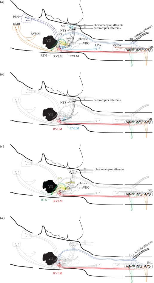
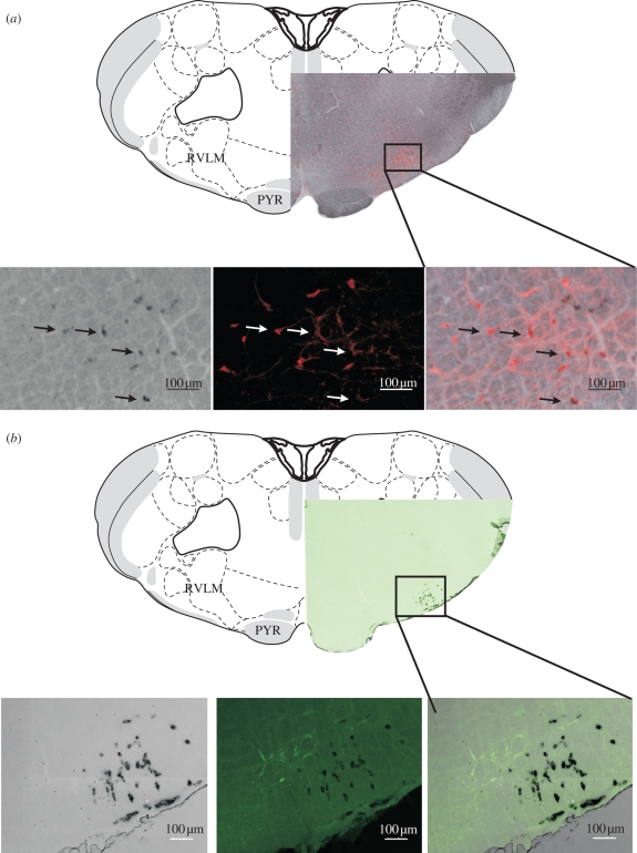
Central neurons in the brainstem and spinal cord are essential for the maintenance of sympathetic tone, the integration of responses to the activation of reflexes and central commands, and the generation of an appropriate respiratory motor output. Here, we will discuss work that aims to understand the role that metabotropic neurotransmitter systems play in central cardiorespiratory mechanisms. It is well known that blockade of glutamatergic, gamma-aminobutyric acidergic and glycinergic pathways causes major or even complete disruption of cardiorespiratory systems, whereas antagonism of other neurotransmitter systems barely affects circulation or ventilation. Despite the lack of an 'all-or-none' role for metabotropic neurotransmitters, they are nevertheless significant in modulating the effects of central command and peripheral adaptive reflexes. Finally, we propose that a likely explanation for the plethora of neurotransmitters and their receptors on cardiorespiratory neurons is to enable differential regulation of outputs in response to reflex inputs, while at the same time maintaining a tonic level of sympathetic activity that supports those organs that significantly autoregulate their blood supply, such as the heart, brain, retina and kidney. Such an explanation of the data now available enables the generation of many new testable hypotheses.


Figures

Figure 1.
Figure 1.
A diagram of pathways in the regulation of the cardiorespiratory system: (a) all pathways overlapped. The bulbospinal red pathways are in the RVLM (figure 2a) and integrate information from the centre and the periphery. The output from this nucleus is crucial for maintaining normal sympathetic tone. PBN, parabrachial nucleus; DMH, dorsomedial hypothalamus; CVLM, caudal ventrolateral medulla; VLM, ventrolateral medulla; rVRG, rostral ventral respiratory group; CPA, caudal pressor area; MCPA, medullo cervical pressor area; IML, intermediolateral cell column; RVMM, rostral ventromedial medulla; VII, facial nucleus; RTN, retrotrapezoid nucleus; preBöt, preBötzinger neurons; VN, vestibular nucleus. (b) The baroreflex pathway is shown on its own. Stretch receptor afferent neurons from the aortic arch and carotid sinus and the neurons synapse in the nucleus tractus solitarius (NTS). Neurons in the NTS then activate inhibitory neurons (blue) in the caudal ventrolateral medulla, which in turn inhibit the neurons in the RVLM; this intense gamma-aminobutyric acid (GABA)-mediated inhibition inhibits sympathetic outflow, causing blood pressure and sympathetic nerve activity to fall. Note also the yellow respiratory neurons that modulate the activity of the cardiovascular neurons (also in c). (c) The pathways for peripheral and central chemoreceptors are shown. Central chemoreceptors are highly responsive to changes in CO2 and are found in the retrotrapezoid nucleus. Many of these chemosensitive neurons (greater than 40%) are galaninergic and Phox2b positive, but all lack tyrosine hydroxylase (Stornetta et al. 2009; figure 2b). Peripheral chemoreception emanates from the carotid body. Neurons terminate in the medial NTS (like the baroreceptors). From here, the excitatory information passes to both respiratory and cardiovascular neurons. (d) The somatosympathetic pathway is shown in an abbreviated form. Afferent nociceptive pathways enter the spinal cord in the dorsal roots, activate circuits locally, and at several stations throughout the neuraxis including the RVLM. This pathway is excitatory and results in the appearance of a variable number of peaks in sympathetic nerve activity, depending on which nerve is recorded from. In the case of the greater splanchnic nerve, this is generally two peaks.
Figure 2.
Figure 2.
Neurotransmitter phenotypes in the RVLM (PYR, pyramidal tract). (a) Neurons in the RVLM that express PNMT (in situ hybridization—black) and are also immunofluorescent for tyrosine hydroxylase (red). The third panel shows a merged image and demonstrates colocalization in many of the cells. The drawing is taken from map 59 of Swanson (1998). Note that the pyramidal tract is present but the olivary nucleus is absent. Note also that both the rostral pole of the nucleus ambiguus and the caudal pole of the facial nucleus are present. These ventral landmarks define the rostrocaudal location of the RVLM (as indicated by the boxed area). (b) Galanin (pre-progalanin-expressing—black) neurons in the retrotrapezoid nucleus are close to and partly intermingled with tyrosine hydroxylase (green) immunofluorescent neurons in the RVLM.

References

    1. Achour L., Labbe-Jullie C., Scott M. G. H., Marullo S.2008An escort for GPCRs: implications for regulation of receptor density at the cell surface. Trends Pharmacol. Sci. 29, 528–535 (doi:10.1016/j.tips.2008.07.009) - DOI - PubMed
    1. Adrian E. D., Bronk D. W., Phillips G.1932Discharges in mammalian sympathetic nerves. J. Physiol. 74, 115–133 - PMC - PubMed
    1. Agnati L. F., Fuxe K., Zoli M., Zini I., Härfstrand A., Toffano G., Goldstein M.1988Morphometrical and microdensitometrical studies on phenylethanolamine-N-methyltransferase- and neuropeptide Y-immunoreactive neurons in the rostral medulla oblongata of the adult and old male rat. Neuroscience 26, 461–478 (doi:10.1016/0306-4522(88)90162-5) - DOI - PubMed
    1. Aicher S. A., Schreihofer A. M., Kraus J. A., Sharma S., Milner T. A., Guyenet P. G.2001Mu-opioid receptors are present in functionally identified sympathoexcitatory neurons in the rat rostral ventrolateral medulla. J. Comp. Neurol. 433, 34–47 (doi:10.1002/cne.1123) - DOI - PubMed
    1. Aicher S. A., Sharma S., Mitchell J. L.2002Co-localization of AMPA receptor subunits in the nucleus of the solitary tract in the rat. Brain Res. 958, 454–458 (doi:10.1016/S0006-8993(02)03693-4) - DOI - PubMed

Publication types

MeSH terms