Review
. 2009 Sep 12;364(1529):2577-87.
doi: 10.1098/rstb.2009.0081.

Structural and functional architecture of respiratory networks in the mammalian brainstem

Affiliations
Review

Structural and functional architecture of respiratory networks in the mammalian brainstem

Jeffrey C Smith et al. Philos Trans R Soc Lond B Biol Sci. .

Abstract

Neural circuits controlling breathing in mammals are organized within serially arrayed and functionally interacting brainstem compartments extending from the pons to the lower medulla. The core circuit components that constitute the neural machinery for generating respiratory rhythm and shaping inspiratory and expiratory motor patterns are distributed among three adjacent structural compartments in the ventrolateral medulla: the Bötzinger complex (BötC), pre-Bötzinger complex (pre-BötC) and rostral ventral respiratory group (rVRG). The respiratory rhythm and inspiratory-expiratory patterns emerge from dynamic interactions between: (i) excitatory neuron populations in the pre-BötC and rVRG active during inspiration that form inspiratory motor output; (ii) inhibitory neuron populations in the pre-BötC that provide inspiratory inhibition within the network; and (iii) inhibitory populations in the BötC active during expiration that generate expiratory inhibition. Network interactions within these compartments along with intrinsic rhythmogenic properties of pre-BötC neurons form a hierarchy of multiple oscillatory mechanisms. The functional expression of these mechanisms is controlled by multiple drives from more rostral brainstem components, including the retrotrapezoid nucleus and pons, which regulate the dynamic behaviour of the core circuitry. The emerging view is that the brainstem respiratory network has rhythmogenic capabilities at multiple hierarchical levels, which allows flexible, state-dependent expression of different rhythmogenic mechanisms under different physiological and metabolic conditions and enables a wide repertoire of respiratory behaviours.


Figures
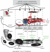
Figure 1.
Figure 1.
Spatially arrayed respiratory compartments extending in the brainstem from the pons to caudal areas of the medulla are illustrated in a schematic parasagittal brainstem section at the level of nucleus ambiguus (NA), VRC, facial nucleus (VII) and the pons, including rostral dorsolateral pontine respiratory regions (PRG consisting of LPBr, lateral parabrachial region and KF, Kölliker–Fuse nucleus). The top schematic illustrates concentrations of excitatory neurons (red circles) and inhibitory neurons (blue circles) in different compartments (cVRG, rVRG, pre-BötC, BötC, RTN/pFRG and PRG). The schematic at the bottom presents a simplified structural view of the serially arranged medullary and pontine compartments, also indicating distributions of the main populations of expiratory and inspiratory neurons within the medullary VRC compartments. Excitatory pre-BötC neurons (top schematic) project (white on red arrows) to rVRG excitatory inspiratory bulbospinal neurons and via premotor circuits to cranial motoneurons (brown circles, hypoglossal XII motoneurons illustrated dorsally). Excitatory expiratory bulbospinal neurons of the cVRG project to thoracic and abdominal spinal expiratory motoneurons. See text for descriptions of functional properties of each compartment and roles of the different populations of excitatory/inhibitory neurons. V, motor nucleus of the trigeminal nerve; Pn, ventral pontine nucleus; LRt, lateral reticular nucleus; SO, superior olive.
Figure 2.
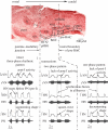
Figure 2.
Transformations of respiratory rhythm and pattern following sequential brainstem transection in the in situ arterially perfused brainstem–spinal cord preparation from juvenile rat, revealing three rhythmic states of the respiratory network as the circuitry is progressively reduced. Top: parasagittal section (Neutral Red stain) of the mature rat brainstem (NAc, compact and semi-compact subdivisions of NA, both within outlined region; V, trigeminal motor nucleus; VII, facial nucleus; 7n, facial nerve; scp, superior cerebellar peduncle; SO, superior olive). Dimensions indicated are typical for a four- to five-week-old rat. Bottom: representative activity patterns of phrenic (PN), hypoglossal (HN) and central vagus (cVN) nerves recorded from the intact preparation (left) and from reduced preparations obtained by transections at the pontine–medullary junction performed to remove the pons (vertical dot-dashed line 1, middle panel) and at the rostral boundary of the pre-BötC made to remove all compartments rostral to pre-BötC (vertical dot-dashed line 2, right panel). Each panel shows raw (bottom traces) and integrated (upper traces) recordings of motor nerve discharge. Vertical dashed lines in the left panel indicate onsets of HN inspiratory burst and the post-I component of cVN activity characteristic of the intact three-phase rhythmic pattern, which also includes a ramping PN discharge. Dashed lines in middle and right panels indicate synchronous onset of inspiratory bursts in all nerves characteristic of the two-phase and one-phase rhythmic patterns. Motor nerve discharges have square-wave and decrementing shapes in the two-phase and one-phase patterns, respectively, which also characterize these rhythmic states (after Smith et al. 2007).
Figure 3.
Figure 3.
Schematic of the core microcircuitry within BötC and pre-BötC respiratory compartments and activity patterns of respiratory neuron populations of the core circuitry. Shown are the main interacting neuronal populations in these compartments operating under the control of external excitatory drives from the pons, RTN and raphé nuclei. Spheres represent neuronal populations (excitatory, red, including tonic drives to the different populations; inhibitory, blue). Red arrow, excitatory connection; small blue circles, inhibitory connection. Inhibitory neurons distributed in the BötC and pre-BötC compartments are interconnected in a mutual inhibitory ring-like circuit (blue background shading, see bottom for activity patterns) that dynamically interacts with the excitatory kernel network in the pre-BötC. The excitatory (glutamatergic) pre-BötC neurons with pre-I/inspiratory discharge patterns have persistent sodium current (INaP) that underlies an important cellular-based oscillatory mechanism contributing to the autorhythmic properties of the pre-BötC network when this structure is isolated and generates a one-phase rhythmic inspiratory pattern. Pre-BötC excitatory neurons rhythmically excite neurons of the rVRG that generate a ramping pattern of excitatory activity (ramp-I) that then drives inspiratory spinal motoneurons such as phrenic motoneurons in the cervical spinal cord. Respiratory neuron population activity patterns, shown below by the colour-filled activity profiles representing integrated population activity (population spike frequency histograms, units are spikes s−1 per neuron), are obtained from a computational model of the network that reproduces the patterns of activity recorded from neuron populations in the BötC, pre-BötC and rVRG compartments (see Smith et al. 2007 for details of the computational model and electrophysiologically recorded activity patterns of the different neuronal populations). The three phases of the respiratory cycle—inspiration (I), and the two phases of expiration, P-I and stage 2 of expiration (E-2)—are indicated. Neurons with peak activity during post-I define this phase (although post-I neuron population activity also typically extends into the next expiratory phase), while neurons with augmenting (aug-E) discharge patterns define E-2. See text for additional descriptions.
Figure 4.
Figure 4.
Schematic of an extended model of the brainstem respiratory network indicating possible state-dependent neuronal interactions between the RTN/pFRG, the core BötC and pre-BötC circuitry and more caudal compartmentalized circuit components. The schematic shows interacting neuronal populations within the major brainstem respiratory compartments (pons, BötC, pre-BötC, rVRG and cVRG) and includes proposed interactions of a late-E neuronal population in the RTN/pFRG, activated by hypercapnia, with the other respiratory neuron populations. The late-E population in the RTN/pFRG receives a necessary excitatory drive from the pons, which under quiet eupnoeic breathing conditions is not sufficient to evoke rhythmic activity in this population. We propose that during quiet breathing, these neurons are not rhythmically active and are inhibited by early-I inhibitory neurons located in the pre-BötC (see connection labelled inspiratory inhibition). Hypercapnia excites late-E neurons in the RTN/pFRG, so that these cells may start generating bursts in advance of bursts of the pre-BötC pre-I/I population. This expressed RTN/pFRG late-E activity excites abdominal expiratory motoneurons possibly via the excitatory bulbospinal premotor neurons located in cVRG as indicated, resulting in late-E activity in abdominal motor nerves (AbN). We also suggest, as indicated in the schematic, that the late-E population excites the pre-I/I population in the pre-BötC, contributing to an earlier onset and enhancement of the pre-I component of hypoglossal nerve (HN) discharge. Thus this schematic incorporates the concept that cells in the RTN/pFRG exhibit both CO2-sensitive tonic and rhythmic activity profiles, the rhythmic cells are also controlled by pontine inputs and these cells have excitatory actions on both inspiratory (including early-I) and expiratory neuronal components of the circuitry. Other cell populations that provide tonic excitatory drive to the network, such as raphé serotonergic neurons, have also been proposed to provide state-dependent excitatory drive to the network, including in response to alterations in blood/brain CO2, as depicted in the schematic. Red arrow, excitatory connection; small blue circles, inhibitory connection; red sphere, excitatory population; blue sphere, inhibitory population; brown sphere, motoneurons.

References

    1. Abdala A. P. L., Rybak I. A., Smith J. C., Paton J. F. R.In press Abdominal expiratory activity in the rat brainstem–spinal cord in situ: patterns, origins, and implications for respiratory rhythm generation. J. Physiol - PMC - PubMed
    1. Alheid G. F., McCrimmon D. R.2008The chemical neuroanatomy of breathing. Respir. Physiol. Neurobiol. 164, 3–11 (doi:10.1016/j.resp.2008.07.014) - DOI - PMC - PubMed
    1. Alheid G. F., Milsom W. K., McCrimmon D. R.2004Pontine influences on breathing: an overview. Respir. Physiol. Neurobiol. 143, 105–114 (doi:10.1016/j.resp.2004.06.016) - DOI - PubMed
    1. Bianchi A. L., Denavit-Saubie M., Champagnat J.1995Central control of breathing in mammals: neuronal circuitry, membrane properties, and neurotransmitters. Physiol. Rev. 75, 1–45 - PubMed
    1. Butera R. J., Rinzel J. R., Smith J. C.1999aModels of respiratory rhythm generation in the pre-Bötzinger complex. I. Bursting pacemaker neurons. J. Neurophysiol. 82, 382–397 - PubMed

Publication types

LinkOut - more resources