. 2009 Sep 15;587(Pt 18):4481-95.
doi: 10.1113/jphysiol.2009.173344. Epub 2009 Aug 3.

GABA blockade unmasks an OFF response in ON direction selective ganglion cells in the mammalian retina

Affiliations

GABA blockade unmasks an OFF response in ON direction selective ganglion cells in the mammalian retina

Jessica M Ackert et al. J Physiol. .

Abstract

One unique subtype of retinal ganglion cell is the direction selective (DS) cell, which responds vigorously to stimulus movement in a preferred direction, but weakly to movement in the opposite or null direction. Here we show that the application of the GABA receptor blocker picrotoxin unmasks a robust excitatory OFF response in ON DS ganglion cells. Similar to the characteristic ON response of ON DS cells, the masked OFF response is also direction selective, but its preferred direction is opposite to that of the ON component. Given that the OFF response is unmasked with picrotoxin, its direction selectivity cannot be generated by a GABAergic mechanism. Alternatively, we find that the direction selectivity of the OFF response is blocked by cholinergic drugs, suggesting that acetylcholine release from presynaptic starburst amacrine cells is crucial for its generation. Finally, we find that the OFF response is abolished by application of a gap junction blocker, suggesting that it arises from electrical synapses between ON DS and polyaxonal amacrine cells. Our results suggest a novel role for gap junctions in mixing excitatory ON and OFF signals at the ganglion cell level. We propose that OFF inputs to ON DS cells are normally masked by a GABAergic inhibition, but are unmasked under certain stimulus conditions to mediate optokinetic signals in the brain.


Figures
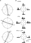
Figure 4
Figure 4. The OFF response of ON DS ganglion cells is direction selective
A, polar plot summarizing the directions for which the ON response of 22 ON DS cells were selective under control conditions and the OFF response unmasked by PTX. The stimulus was a 50 μm wide/275 μm long rectangular slit of light moved in 8 directions. Cells could be divided into three groups in which the ON response was selective roughly to stimulus movement in the dorsal, ventral, or nasal directions. Arrows indicate average summed vector. Grey arrows indicate that summed vectors of directional preference for individual cells, whereas the black arrows indicate the average summed vector for each cell population. Clearly, the OFF response showed a directional preference opposite to that of the ON response for the 3 ON DS cell subtypes. B, responses of a ON DS cell to a 50 μm wide slit of light swept across the retina in 4 directions: dorsal (D), ventral (V), nasal (N), and temporal (T) under control conditions. The cell was direction selective for temporal-to-nasal (leftward) movement along the horizontal axis. After application of PTX, both ON and OFF responses were visible. However, while the direction selectivity of the ON response was abolished by PTX (50 μm), the OFF response showed direction selectivity, but to movement in the nasal-to-temporal (rightward) direction. Application of PTX (50 μm) and l-AP4 (50 μm) abolished the ON response and isolated the OFF response. Slit speed was approximately 180 μm s−1.
Figure 1
Figure 1. Morphology and tracer-coupling pattern of ON DS ganglion cells
A, drawing of a Neurobiotin-labelled ON DS ganglion cell (black) and the somata of tracer-coupled amacrine cells (red). The morphology of one coupled amacrine cell is drawn in detail (blue). Long, straight axon-like processes can be seen extending beyond the field of view (AC axons). In addition, the amacrine cells maintain curvy dendritic processes that end proximally. Scale bar, 25 μm. These morphological features indicate that the amacrine cells are polyaxonal cells with dendritic and axonal systems. B, confocal vertical section showing the level of dendritic stratification of ON DS cell dendrites. Immunolabelling for ChAT (green) shows the two bands of starburst amacrine cell processes defining sublaminae-a and -b of the IPL. The dendrites of the ON DS cell monostratify along the ChAT band in sublamina-b.
Figure 2
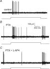
Figure 2. Application of PTX unmasks an OFF response in ON DS ganglion cells
A, typical response of an ON DS cell to a focal light stimulus (275 μm diameter) under control superfusate conditions. Trace below record shows onset and offset of the light stimulus. B, application of PTX (50 μm) reveals a robust OFF response at stimulus offset. C, application of PTX (50 μm) and l-AP4 (50 μm) blocks the ON response, but not the OFF response, indicating that the latter is generated by the OFF retinal pathway.
Figure 3
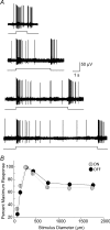
Figure 3. Latencies and area summations of the ON and OFF response of ON DS ganglion cells
A, responses of an ON DS cell after application of PTX to unmask the OFF response. Onset and offset of the full-field light stimulus is shown below each record. Descending down the figure, the response of the cell is shown to stimuli with increasing duration of 1.5, 4.5, 7.0 and 11.0 s. The OFF response always occurs at light offset, irrespective of the stimulus duration, indicating that its latency is not linked to the time of stimulus onset. This result is consistent with idea that the unmasked OFF response is generated by the OFF retinal pathway. B, area summation profiles of the ON and OFF responses to concentric spots of light of increasing diameters (75, 150, 275, 425, 750, 1250 and 1750 μm), but constant luminance (2.5 Rh* rod−1 s−1). Each point is the average of 5 cells; bars indicate standard errors of the mean.
Figure 5
Figure 5. Direction selectivity ratios of the ON and OFF responses of ON DS ganglion cells
A, bar graph showing the average direction selectivity ratio of ON DS cells under control conditions. These cells show a clear direction selectivity along the preferred/null axis for stimulus movement, but no selectivity along the orthogonal axis. Error bars indicate standard errors of the mean. B, left, bar graph showing the average selectivity ratio for the ON response of the same cohort of cells as in A during PTX application. PTX clearly abolishes the direction selectivity of the ON response. B, right, bar graph showing the average selectivity ratio for the OFF response of the same cohort of ON DS cells unmasked by PTX. The selectivity ratio of the OFF response is comparable to that shown by the ON response for stimulus movement along the preferred/null axis. As for the ON response, the OFF response shows no significant direction selectivity along the orthogonal axis.
Figure 6
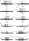
Figure 6. Effect of cholinergic drugs on the direction selectivity of the OFF response of ON DS ganglion cells
A, ON response of an ON DS cell to a 50 μm wide slit moving along the horizontal axis. The cell prefers movement in the temporal to nasal direction. Application of PTX (50 μm) and l-AP4 (50 μm) unmasks and isolates the OFF response in the ON DS cell. The OFF response shows direction selectivity to the moving slit of light, but its preferred direction is nasal to temporal, opposite of that of the ON response. The addition of hexamethonium (Hex) abolishes the direction selectivity of the OFF response, resulting in similar responses to stimulus movement in either direction along the horizontal axis. Slit speed was approximately 80 μm s−1. B, ON response of another ON DS cell to a 50 μm wide slit moving along the vertical axis. The cell prefers movement in the upward direction. Application of PTX (50 μm) and l-AP4 (50 μm) unmasks and isolates the OFF response in the cell. The OFF response shows direction selectivity to the moving slit of light, but its preferred direction is downward, opposite of that of the ON response. The addition of neostigmine (Neo) abolishes the direction selectivity of the OFF response, resulting in similar responses to stimulus movement in either direction along the horizontal axis. Slit speed was approximately 100 μm s−1.
Figure 8
Figure 8. 18β-Glycyrrhetinic acid eliminates the OFF response in ON DS ganglion cells to a moving light stimulus
A, ON response of an ON DS cell to a 50 μm wide slit moving along the vertical axis at approximately 180 μm s−1. The cell prefers movement in the downward direction. B, application of PTX (50 μm) and l-AP4 (50 μm) unmasks and isolates the OFF response in the ON DS cell. The OFF response shows direction selectivity to the moving slit of light, but its preferred direction is upward. C, application of 18β-GA completely eliminates the OFF response to the moving slit of light. D, the direction selective OFF response is completely recovered after washout of 18β-GA.
Figure 7
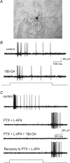
Figure 7. 18β-Glycyrrhetinic acid eliminates the OFF response in ON DS ganglion cells to a stationary light stimulus
A, photomicrograph of an ON DS cell labelled with Neurobiotin after 20 min incubation in 18β-glycyrrhetinic acid (18β-GA; 25 μm). Tracer coupling to neighbouring amacrine cells is completely eliminated. Scale bar, 100 μm. B, response of an ON DS cell to full-field illumination under control conditions. Subsequent application of the gap junction blocker has no effect on the ON response of the ON DS cell. Light trace below shows onset and offset of the light stimulus. C, response of an ON DS cell to full-field illumination under control superfusate conditions. Subsequent application of PTX (50 μm) and l-AP4 (50 μm) unmasks an OFF response and eliminates the ON response. Application of 18β-GA (25 μm) completely abolishes the OFF response. The effect of 18β-GA is reversed upon washout. The onset and offset of the full-field light stimulus are shown by the light trace at bottom of the panel.
Figure 9
Figure 9. Amacrine cells coupled to ON DS ganglion cells cofasciculate with OFF bipolar and starburst amacrine cell dendrites in sublamina-a of the IPL
A, a 1 μm-thick confocal section showing somata of polyaxonal amacrine cells (red) in the proximal INL that are coupled to a Neurobiotin-injected ON DS ganglion cell. The somata of starburst-a amacrine cells are also visible (green) following immunolabelling for ChAT. Scale bar, 20 μm. B, dendrites of polyaxonal amacrine cells cofasciculate with the synaptic ribbons of OFF bipolar cell axon terminals immunolabelled for Ribeye in sublamina-a of the IPL. Scale bar, 20 μm. C, higher magnification of the micrograph in B. Scale bar, 10 μm. D, dendrites of polyaxonal amacrine cells (red) cofasciculate with the dendrites of starburst-a amacrine cells in sublamina-a of the IPL. Scale bar, 20 μm. E, magnified view of the micrograph in D. Scale bar, 10 μm. F, dendrites (arrowheads) of polyaxonal amacrine cells then descend to sublamina-b and costratify with dendrites of ON DS ganglion cell (arrow) and dendrites of starburst-b amacrine cells (green). Scale bar, 20 μm.

References

    1. Ackert JM, Wu SH, Abrams J, Hu EH, Perlman I, Bloomfield SA. Light-induced changes in spike synchronization between coupled ON direction selective ganglion cells in the mammalian retina. J Neurosci. 2006;26:4206–4215. - PMC - PubMed
    1. Ariel M, Daw NW. Pharmacological analysis of directionally sensitive rabbit retinal ganglion cells. J Physiol. 1982;324:161–185. - PMC - PubMed
    1. Barlow HB, Hill RM, Levick WR. Retinal ganglion cells responding selectively to direction and speed of image motion in the rabbit. J Physiol. 1964;173:377–407. - PMC - PubMed
    1. Barlow HB, Levick WR. The mechanism of directionally selective units in the rabbit's retina. J Physiol. 1965;178:477–504. - PMC - PubMed
    1. Bloomfield SA, Miller RF. A functional organization of ON and OFF pathways in the rabbit retina. J Neurosci. 1986;6:1–13. - PMC - PubMed

Publication types

MeSH terms

Substances

LinkOut - more resources