. 2009 Oct 2;284(40):27533-43.
doi: 10.1074/jbc.M109.048884. Epub 2009 Aug 3.

Wnt-5a-induced phosphorylation of DARPP-32 inhibits breast cancer cell migration in a CREB-dependent manner

Affiliations

Wnt-5a-induced phosphorylation of DARPP-32 inhibits breast cancer cell migration in a CREB-dependent manner

Christian Hansen et al. J Biol Chem. .

Abstract

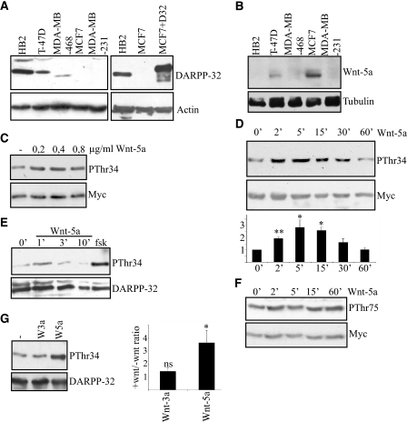
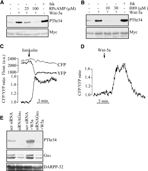
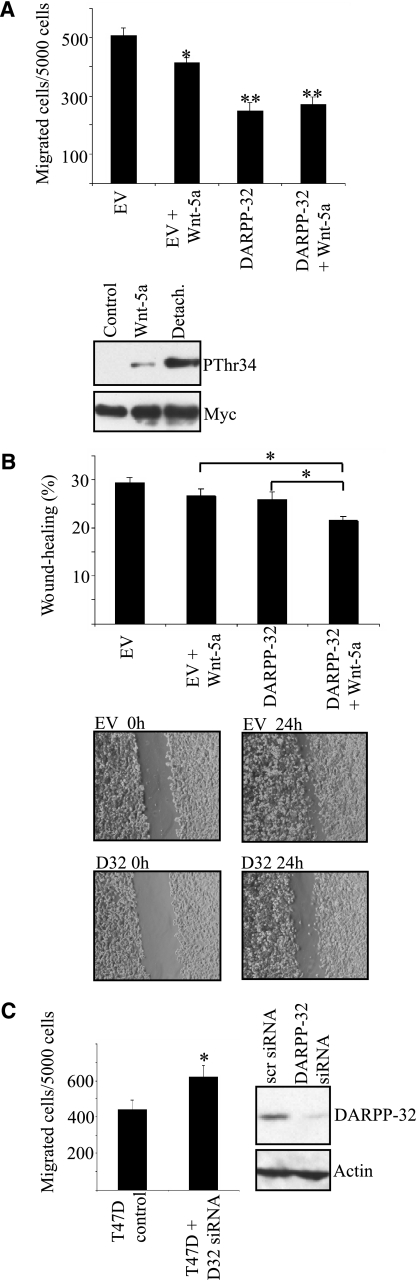
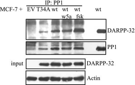
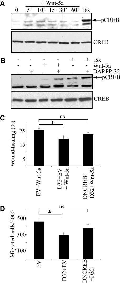
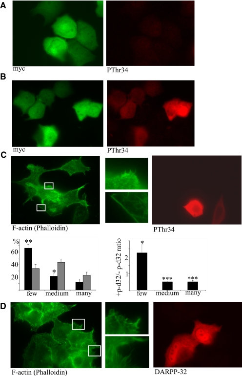
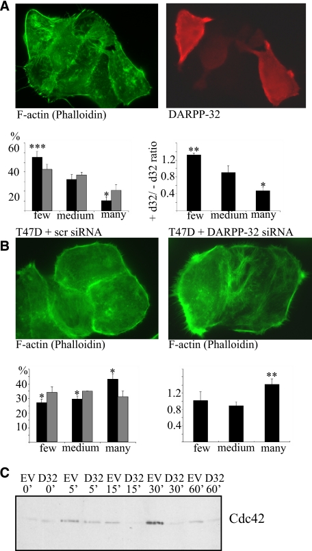
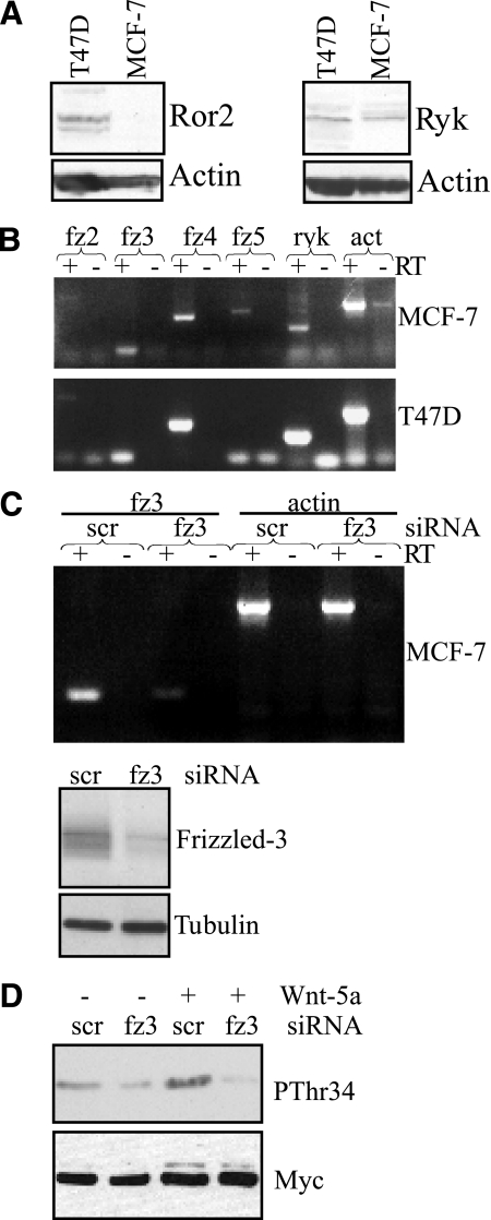
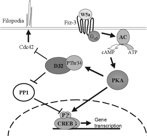
Tumor cell migration plays a central role in the process of cancer metastasis. We recently identified dopamine and cAMP-regulated phosphoprotein of 32 kDa (DARPP-32) as an antimigratory phosphoprotein in breast cancer cells. Here we link this effect of DARPP-32 to Wnt-5a signaling by demonstrating that recombinant Wnt-5a triggers cAMP elevation at the plasma membrane and Thr34-DARPP-32 phosphorylation in MCF-7 cells. In agreement, both protein kinase A (PKA) inhibitors and siRNA-mediated knockdown of Frizzled-3 receptor or Galpha(s) expression abolished Wnt-5a-induced phosphorylation of DARPP-32. Furthermore, Wnt-5a induced DARPP-32-dependent inhibition of MCF-7 cell migration. Phospho-Thr-34-DARPP-32 interacted with protein phosphatase-1 (PP1) and potentiated the Wnt-5a-mediated phosphorylation of CREB, a well-known PP1 substrate, but had no effect on CREB phosphorylation by itself. Moreover, inhibition of the Wnt-5a/DARPP-32/CREB pathway, by expression of dominant negative CREB (DN-CREB), diminished the antimigratory effect of Wnt-5a-induced phospho-Thr-34-DARPP-32. Phalloidin-staining revealed that that the presence of phospho-Thr-34-DARPP-32 in MCF-7 cells results in reduced filopodia formation. In accordance, the activity of the Rho GTPase Cdc42, known to be crucial for filopodia formation, was reduced in MCF-7 cells expressing phospho-Thr-34-DARPP-32. The effects of DARPP-32 on cell migration and filopodia formation could be reversed in T47D breast cancer cells that were depleted of their endogenous DARPP-32 by siRNA targeting. Consequently, Wnt-5a activates a Frizzled-3/Galpha(s)/cAMP/PKA signaling pathway that triggers a DARPP-32- and CREB-dependent antimigratory response in breast cancer cells, representing a novel mechanism whereby Wnt-5a can inhibit breast cancer cell migration.


Figures

FIGURE 1.
FIGURE 1.
Wnt-5a induces Thr-34 phosphorylation of DARPP-32 in MCF-7 and HB2 breast epithelial cells. A, left panel, expression of DARPP-32 in five different breast epithelial cell lines, detected by anti-DARPP-32 Western blotting. Right panel, comparison of endogenous levels of DARPP-32 in HB2 cells to exogenous levels of Myc-tagged DARPP-32 in transfected MCF-7 cells, detected by anti-DARPP-32 Western blotting. B, expression of Wnt-5a in five different breast epithelial cell lines, detected by an anti-Wnt-5a antibody. C, Western blot showing Thr-34 phosphorylation of DARPP-32 in MCF-7 cells expressing Myc-DARPP-32, after 5 min of stimulation with different concentrations of rWnt-5a, as detected by an anti-phospho-Thr-34-DARPP-32 antibody. D, upper panel, Western blot showing the kinetics of Thr-34-DARPP-32 phosphorylation upon stimulation with 0.4 μg/ml rWnt-5a in DARPP-32-expressing MCF-7 cells. Lower panel, densitometric analysis of three different Western blots. The data are given as means ± S.D. E, Western blot showing Thr-34 phosphorylation of endogenous DARPP-32 induced by Wnt-5a in HB2 cells as detected by anti-phospho Thr-34-DARPP-32 antibody. F, Western blot showing the time kinetics of Thr-75 DARPP-32 phosphorylation upon stimulation with 0.4 μg/ml rWnt-5a in Myc-DARPP-32-expressing MCF-7 cells, anti-phospho Thr-75 DARPP-32 antibody. G, left panel, Thr-34-DARPP-32 phosphorylation after 5 min of stimulation with either 0.4 μg/ml rWnt-5a or 0.1 μg/ml rWnt-3a. Right panel, densitometric analysis of five different Western blots. Wnt-3a or Wnt-5a stimulation was measured relative to no stimulation, which was set to 1. All of the Western blots shown are representative of at least three independent experiments.
FIGURE 2.
FIGURE 2.
Wnt-5a-induced Thr-34 phosphorylation of DARPP-32 involves cAMP elevation and activation of PKA. A, Western blot showing that the PKA inhibitor RPcAMPS inhibits rWnt-5a-induced Thr-34-DARPP-32 phosphorylation in DARPP-32-expressing MCF-7 cells, as detected by anti-pThr34 antibody. RPcAMPS was added in serum-free medium 1 h prior to stimulation with 0.4 μg/ml rWnt-5a for 5 min. Stimulation with 1 μm forskolin for 20 min was used as a positive control. B, Western blot showing H89 inhibition of rWnt-5a-induced pThr34-DARPP-32 phosphorylation in Myc-DARPP-32-expressing MCF-7 cells, as detected by anti-pThr34 antibody. H89 was added in serum-free medium 1 h prior to stimulation with 0.4 μg/ml rWnt-5a for 5 min. Stimulation with 1 μm forskolin for 20 min was used as a positive control. C and D, evanescent wave microscopy recording of CFP and YFP fluorescence and the cAMP-dependent CFP/YFP ratio from individual MCF-7 cells expressing a fluorescent cAMP biosensor. The cells are stimulated with 10 μm forskolin (C, n = 8) or 0.4 μg/ml Wnt-5a (D, n = 12) and the prestimulatory CFP/YFP ratio was normalized to unity. E, Thr-34-DARPP-32 phosphorylation after 5 min of stimulation with 0.4 μg/ml rWnt-5a in cells treated with siRNA targeting the Gαs protein or a scrambled sequence as a control. All of the Western blots shown are representative of at least three independent experiments.
FIGURE 3.
FIGURE 3.
Wnt-5a induced phosphorylation of DARPP-32 leads to inhibition of cell migration. A, upper panel, migration of MCF-7 cells transfected with Myc-DARPP-32 or empty vector through collagen-coated wells (n = 8). All statistical significances were calculated in comparison with the empty vector control; Lower panel, Western blot showing comparison of rWnt-5a and detachment induced pThr34-phosphorylation, as detected by anti-pThr34 antibody. B, upper panel, wound-healing assay on MCF-7 cells in collagen-coated plates, transfected with either empty vector or Myc-DARPP-32 and left untreated or stimulated with 0.4 μg/ml rWnt-5a for 24 h (n = 12); lower panel, representative photographs from the wound-healing experiments. C, left panel, migration of T47D cells expressing endogenous DARPP-32 or depleted for DARPP-32 by siRNA targeting (n = 6), right panel, DARPP-32 expression level in T47D cells treated with siRNA targeting DARPP-32 or a scrambled control sequence. Both of the Western blots shown are representative of at least three independent experiments.
FIGURE 4.
FIGURE 4.
DARPP-32 interacts with PP1 in MCF-7 cells. Upper panel, co-immunoprecipitation of PP1 and DARPP-32. MCF-7 cells transfected with either empty vector, Myc-T34ADARPP-32, or Myc-DARPP-32 expression plasmid. Binding of PP1 to DARPP-32 was verified by anti-PP1 immunoprecipitation followed by anti-DARPP-32 Western blotting. Lower panel, aliquots of the lysates to be used for immunoprecipitation experiments were tested for Myc-DARPP-32 expression to verify equal DARPP-32 levels in all Myc-DARPP-32-expressing MCF-7 lysates. The Western blot shown is a representative of three independent co-immunoprecipitation experiments.
FIGURE 5.
FIGURE 5.
Involvement of CREB in Wnt-5a- and DARPP-32-mediated inhibition of cell migration. A, Western blot showing rWnt-5a-induced CREB phosphorylation in MCF-7 cells, as detected by anti-pCREB antibody. B, Western blot showing CREB phosphorylation in MCF-7 cells transfected with empty vector or Myc-DARPP-32 expression plasmid, non-stimulated or stimulated with 0.4 μg/ml rWnt-5a for or 1 μm forskolin for 10 min. Each Western blot is representative of six independent experiments. C, wound-healing assay with MCF-7 cells in collagen-coated plates, transfected with either empty vector, Myc-DARPP-32 vector, or both Myc-DARPP-32 and DN-CREB vectors. All cells were stimulated with 0.4 μg/ml rWnt-5a for 24 h (n = 12). D, migration of MCF-7 through collagen-coated wells transfected with empty vector, Myc-DARPP-32 vector, or both Myc-DARPP-32 and DN-CREB vectors (n = 6).
FIGURE 6.
FIGURE 6.
Thr-34 phosphorylation of DARPP-32 leads to inhibition of filopodia formation. MCF-7 cells expressing Myc-DARPP-32 were plated onto collagen-coated glass slides overnight. A, non-stimulated cells or B, cells stimulated with 1 μm forskolin for 20 min were fixed and stained for Myc and pThr34-DARPP-32. C, upper panel, cells expressing Myc-DARPP-32 were stimulated with 1 μm forskolin for 20 min were fixed and stained for F-actin (with FITC-phalloidin) and pThr34-DARPP-32. Lower panel, quantitative analysis of filopodia formation in pThr34-DARPP-32-positive and -negative MCF-7 cells. The left panel gives the percentage of cells in each category (black bars: + pThr34-DARPP-32, gray bars: - pThr34-DARPP-32). The right panel gives the ratio of pThr34-DARPP-32-positive to pThr34-DARPP-32-negative for each category. D, Myc-T34A-DARPP-32 was expressed in MCF-7 cells, and cells were plated onto collagen-coated glass slides overnight, stimulated with 1 μm forskolin for 20 min, fixed, and stained F-actin (with FITC-phalloidin) and DARPP-32 (n = 5 for all experiments in this figure).
FIGURE 7.
FIGURE 7.
Effect of Wnt-5a and involvement of DARPP-32 and CDC42 in filipodia formation. A, upper panel, Myc-DARPP-32 was expressed in MCF-7 cells, that were then plated onto collagen-coated glass slides overnight and subsequently stimulated with 0.4 μg/ml Wnt-5a for 24 h before being fixed and stained for F-actin (with FITC-phalloidin) and DARPP-32. Lower panel, quantitative analysis of filopodia formation in DARPP-32-positive and -negative MCF-7 cells. The left panel gives the percentage of cells in each category (black bars: + DARPP-32, gray bars: - DARPP-32) (n = 5). The right panel gives the ratio of DARPP-32-positive to DARPP-32-negative for each category. B, upper panel, T47D cells were treated with siRNA against DARPP-32 or scrambled control siRNA and stained for F-actin by FITC-phalloidin. Lower panel, quantitative analysis of filopodia formation in DARPP-32 siRNA- or scrambled siRNA-treated T47D cells. The left panel gives the percentage of cells in each category (black bars: DARPP-32 siRNA, gray bars: scrambled siRNA) (n = 5). The right panel gives the ratio of DARPP-32 siRNA to scrambled siRNA for each category. C, MCF-7 cells transfected with either Myc-DARPP-32 expression vector or empty vector were detached and re-plated onto collagen-coated plates for 0–60 min. Cells were lysed, and active Cdc42 was precipitated in a GST-PAK1 pull-down assay. A representative anti-Cdc42 Western blot of these pulldowns (n = 5) is shown.
FIGURE 8.
FIGURE 8.
Frizzled-3 mediates Wnt-5a-induced phosphorylation of DARPP-32. A, Western blots showing Ror2 and Ryk expression in MCF-7 and T47D cells. B, RT-PCR expression analysis of Frizzled -2,-3,-4,-5, Ryk, and actin. For each expression analysis a negative control lacking reverse transcriptase (−RT) has been included. C, upper panel, RT-PCR of Frizzled-3 in MCF-7 cells transfected with either Frizzled-3 or scrambled siRNA. Actin served as a positive control and for each expression analysis a negative control lacking reverse transcriptase (−RT) has been included. Lower panel, knockdown of Frizzled-3 expression in MCF-7 cells verified by Western blot analysis using an anti-Frizzled-3 antibody. D, Western blot analysis showing Wnt-5a (0.4 μg/ml, 5 min)-mediated Thr-34 phosphorylation in DARPP-32-expressing MCF-7 cells co-transfected with either Frizzled-3 siRNA or scrambled siRNA. The Western blots and RT-PCRs shown are representative of at least three independent experiments.
FIGURE 9.
FIGURE 9.
Model of Wnt-5a/DARPP-32-mediated inhibition of breast cancer cell migration. Autocrine or paracrine acting Wnt-5a ligand activates Frizzled-3 receptors that via Gαs stimulates cAMP formation by adenylyl cyclases (AC). cAMP activates PKA, which phosphorylates DARPP-32 at Thr-34 and CREB at Ser-133. pThr34-DARPP-32 suppresses filopodia formation in part by reducing CDC42 activity. In addition, pThr34-DARPP-32 inhibits PP1, which in turn leads to enhanced CREB phosphorylation. CREB mediates transcription of genes that also regulate cell migration by an as yet unidentified mechanism.

References

    1. Hansen C., Greengard P., Nairn A. C., Andersson T., Vogel W. F. (2006) Exp. Cell Res. 312, 4011–4018 - PubMed
    1. Walaas S. I., Aswad D. W., Greengard P. (1983) Nature 301, 69–71 - PubMed
    1. Hemmings H. C., Jr., Greengard P., Tung H. Y., Cohen P. (1984) Nature 310, 503–505 - PubMed
    1. Bibb J. A., Snyder G. L., Nishi A., Yan Z., Meijer L., Fienberg A. A., Tsai L. H., Kwon Y. T., Girault J. A., Czernik A. J., Huganir R. L., Hemmings H. C., Jr., Nairn A. C., Greengard P. (1999) Nature 402, 669–671 - PubMed
    1. Svenningsson P., Nishi A., Fisone G., Girault J. A., Nairn A. C., Greengard P. (2004) Annu. Rev. Pharmacol. Toxicol. 44, 269–296 - PubMed

Publication types

MeSH terms