. 2009 Oct 2;284(40):27123-34.
doi: 10.1074/jbc.M109.051235. Epub 2009 Aug 3.

Inducible nitric-oxide synthase expression is regulated by mitogen-activated protein kinase phosphatase-1

Affiliations

Inducible nitric-oxide synthase expression is regulated by mitogen-activated protein kinase phosphatase-1

Xianxi Wang et al. J Biol Chem. .

Abstract

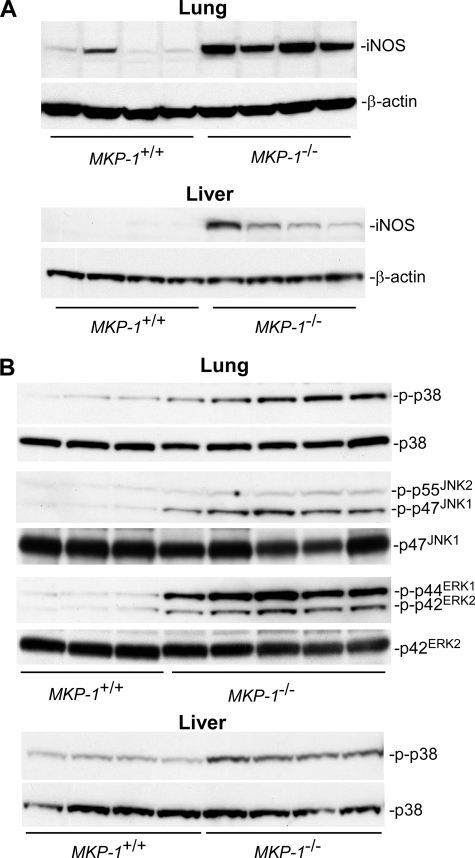
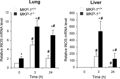
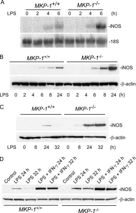
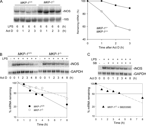
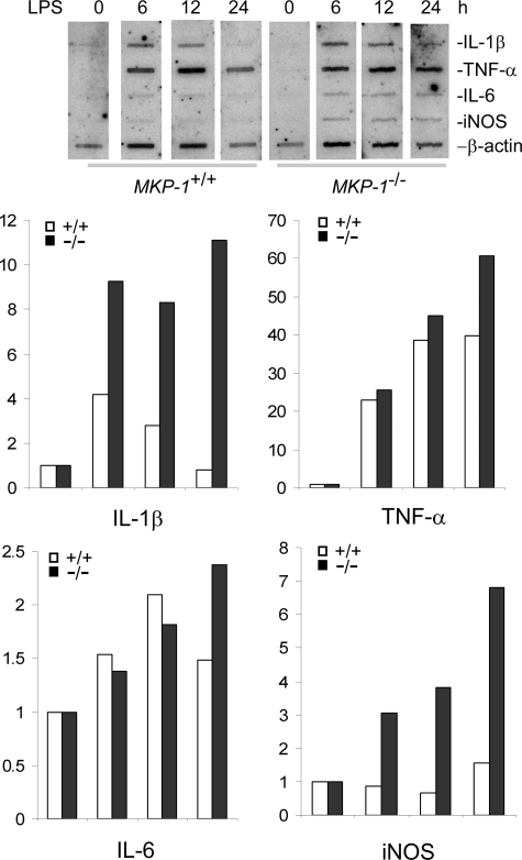
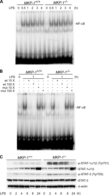
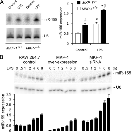
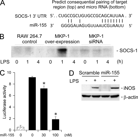
Inducible nitric-oxide (NO) synthase (iNOS) plays a critical role in the eradication of intracellular pathogens. However, the excessive production of NO by iNOS has also been implicated in the pathogenesis of septic shock syndrome. Previously, we have demonstrated that mice deficient in mitogen-activated protein kinase phosphatase-1 (MKP-1) exhibit exaggerated inflammatory responses and rapidly succumb to lipopolysaccharide (LPS). In response to LPS, MKP-1(-/-) mice produce greater amounts of inflammatory cytokines and NO than do wild-type mice, and the MKP-1(-/-) mice exhibit severe hypotension. To understand the molecular basis for the increase in NO production, we studied the role of MKP-1 in the regulation of iNOS expression. We found that LPS challenge elicited a stronger iNOS induction in MKP-1 knock-out mice than in wild-type mice. Likewise, LPS treatment also resulted in greater iNOS expression in macrophages from MKP-1(-/-) mice than in macrophages from wild-type mice. Both accelerated gene transcription and enhanced mRNA stability contribute to the increases in iNOS expression in LPS-stimulated MKP-1(-/-) macrophages. We found that STAT-1, a transcription factor known to mediate iNOS induction by interferon-gamma, was more potently activated by LPS in MKP-1(-/-) macrophages than in wild-type cells. MicroRNA array analysis indicated that microRNA (miR)-155 expression was increased in MKP-1-deficient macrophages compared with wild-type macrophages. Transfection of miR-155 attenuated the expression of Suppressor of Cytokine Signal (SOCS)-1 and enhanced the expression of iNOS. Our results suggest that MKP-1 may negatively regulate iNOS expression by controlling the expression of miR-155 and consequently the STAT pathway via SOCS-1.


Figures

FIGURE 1.
FIGURE 1.
LPS challenge results in enhanced iNOS induction and greater MAP kinase activity in MKP-1−/− mice than in wild-type mice. MKP-1+/+ and MKP-1−/− mice were injected intraperitoneal with LPS (1.5 mg/kg body weight) and sacrificed 3 or 24 h later. Lung and liver tissues were collected, and tissue homogenates were analyzed for the levels of iNOS and phospho-MAP kinases by Western blotting. The membranes were stripped and reblotted with an antibody for β-actin or total MAP kinase as loading controls. A, iNOS protein levels in the lungs and livers of MKP-1+/+ and MKP-1−/− mice at 24 h post-LPS challenge. B, MAP kinase activities in lung and liver homogenates from MKP-1+/+ and MKP-1−/− mice 3 h post-LPS challenge.
FIGURE 2.
FIGURE 2.
MKP-1 knock-out mice exhibit greater iNOS mRNA expression in both lung and liver in response to LPS challenge than do wild-type mice. MKP-1+/+ and MKP-1−/− mice were challenged intraperitoneal with phosphate-buffered saline (0 h) or with LPS (1.5 mg/kg body weight) and sacrificed 3 or 24 h post-challenge. Total RNA was isolated from the lung and liver of these animals. Quantitative real-time PCR was carried out to determine the iNOS mRNA levels in tissues. GAPDH mRNA was used as an internal control for normalization between samples. Values are expressed as -fold increase relative to the iNOS mRNA levels in the lung or liver tissues isolated from vehicle-treated wild-type mice. Data are presented as the mean ± S.E. (n = 3). *, MKP-1−/− different from MKP-1+/+ at same time point, p < 0.05; #, different from vehicle-treated mice of the same genotype, p < 0.05. Left, iNOS mRNA levels in the lungs from MKP-1+/+ and MKP-1−/− mice. Right, iNOS mRNA levels in livers from MKP-1+/+ and MKP-1−/− mice.
FIGURE 3.
FIGURE 3.
LPS stimulation results in greater iNOS induction in MKP-1-deficient peritoneal macrophages than in wild-type cells. A, augmented iNOS mRNA induction by LPS in thioglycollate-elicited MKP-1-deficient peritoneal macrophages. Thioglycollate-elicited peritoneal macrophages were isolated from MKP-1+/+ and MKP-1−/− mice. Cells were treated with 100 ng/ml LPS for the indicated periods. Northern blot analysis was carried out to access the expression of iNOS mRNA. post- ribosomal RNA was used as a loading control. B, more robust iNOS protein increases in MKP-1-deficient macrophages upon LPS stimulation. Thioglycollate-elicited peritoneal macrophages were treated as in A, and Western blotting was carried out to determine levels of iNOS protein. C, the effect of MKP-1 knock-out on iNOS protein induction in resident peritoneal macrophages stimulated with LPS. Resident peritoneal macrophages were stimulated with 100 ng/ml LPS. D, the effect of IFN-γ on iNOS expression in LPS-stimulated wild-type and MKP-1-deficient resident peritoneal macrophages. Cells were treated with 100 ng/ml LPS and 50 IU/ml IFN-γ. iNOS and β-actin levels were assessed by Western blotting. Results from representative experiments are shown.
FIGURE 4.
FIGURE 4.
Knock-out of MKP-1 enhances the stability of iNOS mRNA, and inhibition of p38 decreases iNOS mRNA stability. Thioglycollate-elicited peritoneal macrophages were treated with LPS (100 ng/ml) for 6 h (A), 24 h (B), or 23 h (C), and actinomycin D (5 μg/ml) was added in the medium to inhibit transcription. Total RNA was isolated at the indicated time points after the addition of actinomycin D (Act D). Northern blot analysis was used to determine iNOS mRNA levels. Levels of iNOS mRNA were normalized to 18 S, and the quantified data are presented graphically. A, iNOS mRNA decay assay following 6 h of LPS stimulation. B, iNOS mRNA decay assay after 24 h of LPS stimulation. C, effects of p38 inhibitor on iNOS mRNA stability. MKP-1-deficient peritoneal macrophages were first stimulated with LPS for 23 h, then treated with SB203580 (SB, 10 μm) for 1 h. Actinomycin D was added thereafter and iNOS mRNA decay was examined. The experiments were performed at least three times with similar results. Data presented in the panels are from representative experiments.
FIGURE 5.
FIGURE 5.
Knock-out of MKP-1 enhances iNOS transcription triggered by LPS in primary macrophages. Thioglycollate-elicited peritoneal macrophages isolated from MKP-1+/+ and MKP-1−/− mice were stimulated with LPS (100 ng/ml) for the indicated times, and the nuclei were isolated. Gene transcription was resumed by incubating the nuclei with nucleic acids including [α-32P]UTP to make nascent RNAs as radioactive probes. These probes were then hybridized with membrane-bound mouse cDNAs corresponding to different genes. The signals of given genes were normalized to that of the housekeeping gene β-actin. Data are expressed as -fold induction relative to unstimulated controls (0 h). The experiments were performed twice, with similar results. Data presented are from a representative experiment.
FIGURE 6.
FIGURE 6.
MKP-1 deficiency leads to more robust STAT-1 and STAT-3 tyrosine phosphorylation but has no effect on NF-κB DNA binding activity in primary macrophages stimulated with LPS. A, NF-κB DNA binding activity in wild-type and MKP-1−/− primary macrophages in response to LPS. Nuclear protein was isolated from thioglycollate-elicited peritoneal macrophages stimulated with LPS (100 ng/ml) for the indicated times. A gel shift assay using the 32P-labeled consensus NF-κB oligonucleotide probes was utilized to determine the DNA binding ability of NF-κB. B, specificity of the gel shift assays. Different concentrations of cold wild-type (wt) or mutant (mut) NF-κB oligonucleotides were utilized to compete with the radio-labeled NF-κB probe. C, enhanced STAT-1 and STAT-3 tyrosine phosphorylation upon LPS stimulation in MKP-1-deficient macrophages. Thioglycollate-elicited peritoneal macrophages isolated from MKP-1+/+ and MKP-1−/− mice were treated with 100 ng/ml LPS for the indicated times. Western blotting was carried out to determine levels of STAT-1 (Tyr-701) and STAT-3 (Tyr-705) phosphorylation. The blots were striped and blotted with STAT-1, STAT-3, or β-actin antibody. Data shown are from representative experiments.
FIGURE 7.
FIGURE 7.
MKP-1 negatively regulates the expression of miR-155. A, miR-155 expression in MKP-1+/+ and MKP-1−/− macrophages. Thioglycollate-elicited peritoneal macrophages isolated from MKP-1+/+ and MKP-1−/− mice were stimulated with LPS (100 ng/ml) for 4 h, and total RNA was subjected to Northern blot analysis. U6 small nuclear RNA was used as a loading control for signal normalization. miR-155 expression is presented in the graph on the right as -fold relative to miR-155 expression level in unstimulated wild-type macrophages. The graph on the right represents the means ± S.E. of the relative miR-155 expression calculated from three separate experiments. Relative expression was calculated as (miR-155 valuegiven sample/average miR-155 valueall samples)/(U6 valuegiven sample/average U6 Valueall samples). *, p < 0.05, relative to control; §, p < 0.05, relative to similarly treated wild-type cells. Data were analyzed using a Student's t test. B, effects of MKP-1 overexpression or knock-down on miR-155 induction by LPS in RAW264.7 macrophages. The establishment of RAW264.7 cells harboring an empty vector, a MKP-1 overexpression construct, or a construct expressing a MKP-1 siRNA were previously described (11, 12). These stable clones were stimulated with LPS (100 ng/ml) or left untreated. miR-155 expression was examined by Northern blot analysis. The graph underneath the images represents means ± S.E. of the relative miR-155 expression calculated from three experiments. By two-way ANOVA there was an effect of time (p < 0.05), cell line (p < 0.05) and a statistically significant interaction among these groups (p < 0.05).
FIGURE 8.
FIGURE 8.
miR-155 attenuates the expression of SOCS-1 and enhances the expression of iNOS. A, the miR-155 targeting sequence in the 3′-UTR of murine SOCS-1 mRNA. Paring with miR-155 was also illustrated. B, the effect of MKP-1 overexpression or knockdown on SOCS-1 expression in LPS-treated RAW264.7 macrophages. Stable clones of RAW264.7 macrophages expressing a MKP-1 cDNA, a MKP-1 siRNA, or carrying an empty vector were stimulated with LPS (100 ng/ml) for the indicated periods. SOCS-1 expression was assessed by immunoprecipitation followed by Western blotting. C, effects of miR-155 on a luciferase reporter containing a SOCS-1-derived putative miR-155 targeting sequence. Luciferase reporter containing a SOCS-1-derived putative miR-155 targeting sequence together with an internal control reporter (Renilla luciferase) and different concentrations of miR-155 was transfected into HEK 293 cells. Luciferase activity was measured 48 h post-transfection, and values were normalized to Renilla luciferase activity. Data represent the ratio between the firefly and Renilla luciferase activities. The striped bar represents a background reading from a promoterless firefly luciferase reporter. Data were presented as means ± S.E. of three independent experiments. D, effect of miR-155 transfection on iNOS induction by LPS in RAW264.7 macrophages. RAW264.7 cells were transiently transfected with miR-155 or scramble RNA and then stimulated with LPS (100 ng/ml) 24 h later. iNOS protein levels in these cells were assessed by Western blotting. Data shown are from a representative experiment.

References

    1. Bogdan C. (2001) Nat. Immunol. 2, 907–916 - PubMed
    1. Janeway C. A. J., Travers P., Walport M., Shlomchik M. J. (2005) Immunobiology: The Immune System in Health and Disease, 6th Ed., pp. 37–100, Garland Publishing, New York
    1. Bogdan C., Röllinghoff M., Diefenbach A. (2000) Immunol. Rev. 173, 17–26 - PubMed
    1. Gordon S. (1995) BioEssays 17, 977–986 - PubMed
    1. Malu S., Srinivasan S., Kumar Maiti P., Rajagopal D., John B., Nandi D. (2003) J. Immunol. Methods 272, 55–65 - PubMed

Publication types

MeSH terms