. 2009 Oct 2;284(40):27416-24.
doi: 10.1074/jbc.M109.031278. Epub 2009 Aug 3.

Rapamycin rescues TDP-43 mislocalization and the associated low molecular mass neurofilament instability

Affiliations

Rapamycin rescues TDP-43 mislocalization and the associated low molecular mass neurofilament instability

Antonella Caccamo et al. J Biol Chem. .

Abstract

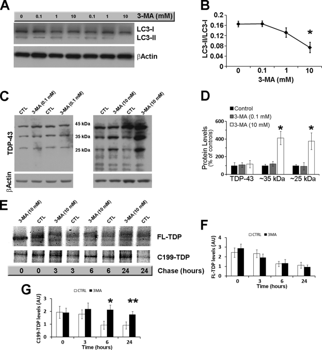
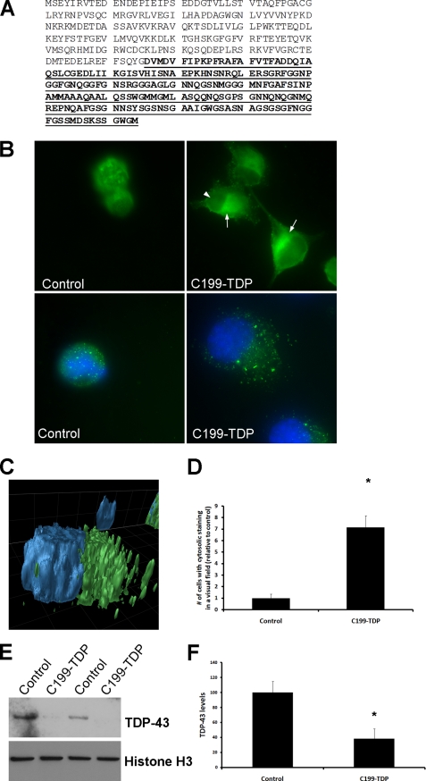
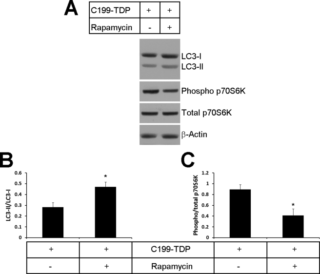
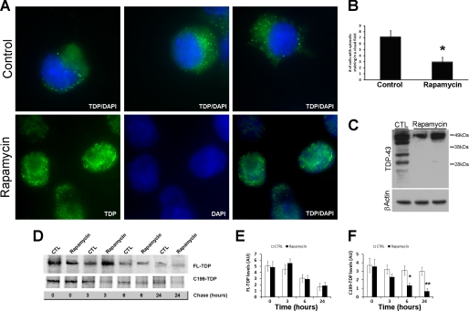
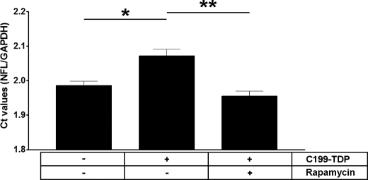
TDP-43 is a nuclear protein involved in exon skipping and alternative splicing. Recently, TDP-43 has been identified as the pathological signature protein in frontotemporal lobar degeneration with ubiquitin-positive inclusions and in amyotrophic lateral sclerosis. In addition, TDP-43-positive inclusions are present in Parkinson disease, dementia with Lewy bodies, and 30% of Alzheimer disease cases. Pathological TDP-43 is redistributed from the nucleus to the cytoplasm, where it accumulates. An approximately 25-kDa C-terminal fragment of TDP-43 accumulates in affected brain regions, suggesting that it may be involved in the disease pathogenesis. Here, we show that overexpression of the 25-kDa C-terminal fragment is sufficient to cause the mislocalization and cytoplasmic accumulation of endogenous full-length TDP-43 in two different cell lines, thus recapitulating a key biochemical characteristic of TDP-43 proteinopathies. We also found that TDP-43 mislocalization is associated with a reduction in the low molecular mass neurofilament mRNA levels. Notably, we show that the autophagic system plays a role in TDP-43 metabolism. Specifically, we found that autophagy inhibition increases the accumulation of the C-terminal fragments of TDP-43, whereas inhibition of mTOR, a key protein kinase involved in autophagy regulation, reduces the 25-kDa C-terminal fragment accumulation and restores TDP-43 localization. Our results suggest that autophagy induction may be a valid therapeutic target for TDP-43 proteinopathies.


Figures

FIGURE 1.
FIGURE 1.
The autophagic system is involved in TDP-43 metabolism. Cells transfected with FL-TDP were treated with different concentrations of the autophagy inhibitor 3-MA or vehicle. A, representative Western blot showing the effects of 3-MA on the autophagy marker LC3. β-Actin was used as a loading control. B, quantitative assessment of the LC3-II/LC3-I ratio, which is a reflection of autophagy induction, shows a dose-dependent decline with a maximum and significant decrease at 10 mm (p = 0.0096). C, representative Western blots probed with anti-TDP-43 monoclonal antibody 2E2-D3. β-Actin was used as loading control. Note the increase in the steady-state levels of low molecular mass TDP-43 fragments after the administration of 10 mm 3-MA (right panel) that was not evident after administration of 0.1 mm 3-MA (left panel). CTL, control. D, quantitative assessment of the percentage changes in FL-TDP and the ∼35- and ∼25-kDa C-terminal fragments after administration of 0.1 and 10 mm 3-MA. *, p < 0.001. E, representative Western blot after pulse-chase experiments. After being pulse-labeled for 2 h, cells were chased for the indicated time points. F and G, quantitative assessment of the FL-TDP and C199-TDP levels after the pulse-chase experiments indicates that the FL-TDP levels decreased similarly in the control and 3-MA-treated cells (p > 0.05). In contrast, the C199-TDP levels were significantly higher in 3-MA-treated cells compared with control cells at 6 and 24 h. *, p < 0.01; **, p < 0.05. Data were analyzed using one-way analysis of variance, followed by the post hoc Bonferroni test to determine individual differences in groups. AU, arbitrary units.
FIGURE 2.
FIGURE 2.
GFP levels are not altered by 3-MA treatment. Proteins extracted from cells double-transfected with FL-TDP and GFP were treated with 3-MA or vehicle as described under “Experimental Procedures.” A, representative Western blot probed with anti-GFP antibody. β-Actin was used as a loading control. B, quantitative assessment of the steady-state levels of GFP indicated. Note that the 3-MA treatment did not significantly affect GFP levels as determined by t test analysis (p > 0.05). CTL, control; A.U., arbitrary units.
FIGURE 3.
FIGURE 3.
C199-TDP is sufficient to induce nuclear TDP-43 depletion and cytoplasmic TDP-43 accumulation. A, amino acid sequence of TDP-43 (NCBI accession number NP_031401). The putative 199-amino acid sequence of the ∼25-kDa fragment is underlined and in boldface. Notably, this fragment has been isolated from FTLD-U and ALS brains. B, representative microphotographs of cells transfected with C199-TDP or vector alone. The arrows point to cytoplasmic TDP-43-positive accumulation. Note the reduction in nuclear staining (arrowhead), which is also evident by reduced TDP-43 localization with 4′,6-diamidino-2-phenylindole. C, three-dimensional reconstruction of a z-stack showing that the TDP-43 immunoreactivity (green) is predominately outside the nucleus (blue). D, quantitative assessment of the number of cells with cytosolic TDP-43 staining after C199-TDP expression. *, p < 0.01 as determined by t test analysis. E, representative Western blot of the nuclear fraction of cells transfected with C199-TDP, further confirming the decrease in endogenous TDP-43 levels in the nucleus. F, quantitative assessment of the Western blots shows an ∼60% decrease in nuclear steady-state levels of TDP-43 after C199-TDP transfection. *, p < 0.01 as determined by t test analysis.
FIGURE 4.
FIGURE 4.
Rapamycin administration increases autophagy by decreasing mTOR activity. After transfection with C199-TDP, cells were treated with rapamycin or vehicle as described under “Experimental Procedures.” A, representative Western blots probed with LC3 (to measure autophagy induction) and phospho-p70S6K and total p70S6K (to measure mTOR activity). β-Actin was used as a loading control. B, quantitative assessment of the Western blots shows that, after rapamycin administration, there was a significant increase in the LC3-II/LC3-I ratio, which indicates an increase in autophagy induction. p = 0.0250 as determined by t test analysis. C, quantitative assessment of the Western blots shows that, after rapamycin administration, there was a significant decrease in p70S6K, which indicates a decrease in mTOR activity. p = 0.0343 as determined by t test analysis.
FIGURE 5.
FIGURE 5.
Rapamycin reduces cytosolic TDP-43 immunoreactivity. Cells transfected with C199-TDP were treated with rapamycin or vehicle as described under “Experimental Procedures.” A, representative microphotographs of cells stained with 2E2-D3. Remarkably, rapamycin administration significantly reduced TDP-43 mislocalization as denoted by the increased TDP-43 immunoreactivity (green) in the nucleus (blue). B, quantitative assessment of the number of cells showing cytosolic TDP-43 immunoreactivity after C199-TDP expression. Rapamycin reduced the number of cells showing cytosolic TDP-43 immunoreactivity by ∼60%. p = 0.0016 as determined by t test analysis. C, representative Western blot probed with anti-TDP-43 monoclonal antibody 2E2-D3 showing a significant decrease in the steady-state levels of the C-terminal fragments of TDP-43. D, representative Western blot after pulse-chase experiments. After being pulse-labeled for 2 h, cells were chased for the indicated time points. E and F, quantitative assessment of the FL-TDP and C199-TDP levels after the pulse-chase experiments indicates that the FL-TDP levels decreased similarly in the control (CTL) and rapamycin-treated cells (p > 0.05). In contrast, C199-TDP levels were significantly lower in rapamycin-treated cells compared with control cells at 6 and 24 h, indicating that rapamycin facilitates the turnover of C199-TDP. *, p < 0.01; **, p < 0.001 as determined using one-way analysis of variance, followed by the post hoc Bonferroni test to determine individual differences in groups. DAPI, 4′,6-diamidino-2-phenylindole; AU, arbitrary units.
FIGURE 6.
FIGURE 6.
Rapamycin administration increases NFL mRNA levels. The mRNA levels of NFL were measured using real-time PCR in cells stably transfected with C199-TDP and treated with rapamycin or vehicle only. The NFL mRNA levels were compared with control cells. We found that expression of C199-TDP was sufficient to reduce the levels of NFL mRNA as indicated by higher Ct values. Notably, rapamycin administration rescued the C199-TDP-induced changes in mRNA levels. *, p < 0.01; **, p < 0.001 as determined using one-way analysis of variance, followed by the post hoc Bonferroni test to determine individual differences in groups. GAPDH, glyceraldehyde-3-phosphate dehydrogenase.

References

    1. Wang H. Y., Wang I. F., Bose J., Shen C. K. (2004) Genomics 83, 130–139 - PubMed
    1. Buratti E., Baralle F. E. (2001) J. Biol. Chem. 276, 36337–36343 - PubMed
    1. Buratti E., Dörk T., Zuccato E., Pagani F., Romano M., Baralle F. E. (2001) EMBO J. 20, 1774–1784 - PMC - PubMed
    1. Wang I. F., Reddy N. M., Shen C. K. (2002) Proc. Natl. Acad. Sci. U.S.A. 99, 13583–13588 - PMC - PubMed
    1. Buratti E., Baralle F. E. (2008) Front. Biosci. 13, 867–878 - PubMed

MeSH terms