. 2009 Sep;37(17):5868-80.
doi: 10.1093/nar/gkp635. Epub 2009 Aug 3.

Novel structural determinants in human SECIS elements modulate the translational recoding of UGA as selenocysteine

Affiliations

Novel structural determinants in human SECIS elements modulate the translational recoding of UGA as selenocysteine

Lynda Latrèche et al. Nucleic Acids Res. 2009 Sep.

Abstract

The selenocysteine insertion sequence (SECIS) element directs the translational recoding of UGA as selenocysteine. In eukaryotes, the SECIS is located downstream of the UGA codon in the 3'-UTR of the selenoprotein mRNA. Despite poor sequence conservation, all SECIS elements form a similar stem-loop structure containing a putative kink-turn motif. We functionally characterized the 26 SECIS elements encoded in the human genome. Surprisingly, the SECIS elements displayed a wide range of UGA recoding activities, spanning several 1000-fold in vivo and several 100-fold in vitro. The difference in activity between a representative strong and weak SECIS element was not explained by differential binding affinity of SECIS binding Protein 2, a limiting factor for selenocysteine incorporation. Using chimeric SECIS molecules, we identified the internal loop and helix 2, which flank the kink-turn motif, as critical determinants of UGA recoding activity. The simultaneous presence of a GC base pair in helix 2 and a U in the 5'-side of the internal loop was a statistically significant predictor of weak recoding activity. Thus, the SECIS contains intrinsic information that modulates selenocysteine incorporation efficiency.


Figures

Figure 1.
Figure 1.
SECIS elements from human selenoproteome. (A) Schematic of eukaryotic SECIS elements. Human type 1 elements include Gpx1, Gpx2, SelN, Dio1 and SelV, whereas the other elements are type 2, containing an additional bulge in the apical loop. The four main structural motifs are identified by arrows. A bracket indicates the position of the SECIS core that contains a quartet of non-Watson–Crick base pairs. Highly conserved nucleotides are represented in letters. The dots symbolize the two sheared tandem GA base pairs that are essential for kink-turn structures. The asterisks indicate the variations from the consensus sequence (AA) that are present as CC in SelM, SelO and Trxr3 SECIS elements. (B) Sequence alignment of the 26 human SECIS elements that were cloned downstream of luciferase UGA258 coding sequence and used in the reporter constructs. They were obtained from SelenoDB website (51). Highly conserved nucleotides are represented in bold. Nucleotides involved in helix 2 are underlined. Gpx, glutathione peroxidase; Trxr, thioredoxin reductase; Dio, iodothyronine deiodinase; Sel, selenoprotein; Sel15, 15 kDa selenoprotein, Sps2, selenophosphate synthetase.
Figure 2.
Figure 2.
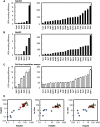
The UGA-selenocysteine recoding efficiency is strongly influenced by the nature of the SECIS element in vivo and in vitro. Luciferase UGA258-SECIS reporter gene constructs were generated with all human SECIS elements and tested in transiently transfected Hek293 (A) or HepG2 (B) cells (black bars), or in the cell free translation system (C, white bars). For in vivo experiments (A and B), each luciferase plasmid was transiently co-transfected with β-galactosidase plasmid. Enzymatic activities were measured 48-h post-transfection on cell protein extracts. Transfection efficiencies were normalized by calculating the ratio between luciferase relative to β-galactosidase activities. For in vitro experiments (C), the luciferase UGA258-SECIS synthetic mRNAs were translated using RRL as described in ‘Materials and Methods’ section. To analyze data from three independent experiments in vivo and in vitro, the UGA recoding efficiencies were arbitrarily expressed relative to the activity from the luciferase UGA258-SelX construct, which was set as 100%. The group of constructs that show weak efficiencies (SelH, SelO, SelS, Gpx3, Gpx6 and Trxr3) are represented at a magnified scale in left panel. (D) Comparison of UGA–selenocysteine recoding efficiencies of our luciferase UGA258-SECIS reporter gene constructs between the different experimental conditions: HepG2 versus Hek293 transfection (left panel), in vitro translation versus Hek293 tranfection (middle panel) and in vitro translation versus HepG2 tranfection (left panel). The SECIS elements are represented as a function of recoding efficiency group according to Figure 6: blue circles (weak), red square (moderate) and yellow triangles (strong).
Figure 3.
Figure 3.
The UGA recoding efficiencies of the SelX and Gpx3 SECIS elements are specific for selenocysteine insertion in vivo and in vitro. (A) The secondary structures of SelX and Gpx3 SECIS elements are represented in the frame of type 2 category of elements. Consensus motifs are shown in bold. The A–U, G–C, G–U and noncanonical G–A base pairs are represented by dots. The main four structural motifs are separated by gray bars. (B) Mutant luciferase-SelX and luciferase-Gpx3 constructs were assayed for UGA recoding activity in transiently transfected Hek293 cells (black bars) or in the rabbit reticulocyte cell-free translation system (white bars). Mutations targeted either essential motifs the SECIS elements (ΔAUGA, ΔAAAC and ΔAAAG) or codon 258 in the luciferase coding sequence (UAA258 and UGU258). UGA recoding activities were expressed relative to the activity of the wild-type luciferase UGA258-SelX construct (set as 100%).
Figure 4.
Figure 4.
SBP2 binds with similar affinity to the SelX (A) and Gpx3 (B) SECIS elements. The 32P-labeled SECIS elements were incubated with increasing concentration of the RNA binding domain of SBP2 (SBP2-RBD). The RNA–protein complexes were analyzed on a native 8% polyacrylamide gel. The apparent Kd values were determined by plotting the fraction of protein–SECIS complex as a function of protein concentration.
Figure 5.
Figure 5.
Structure-function analyses of SelX and Gpx3 SECIS elements. SelX and Gpx3 were used as a representative weak and strong element, respectively. Chimeric constructs of these elements were generated based on the four domains shown in Figure 3A and cloned downstream of luciferase UGA258 coding sequence. The composition and nomenclature of the chimeric SECIS elements are represented beneath histograms, using gray and black to indicate domains originating from Gpx3 and SelX SECIS elements, respectively. The histograms represent data from transiently transfected Hek293 cells (black bars) or from cell free translation assays (white bars), which were performed as described in the Figure 2 caption. The UGA recoding efficiencies are expressed relative to the luciferase UGA258-SelX construct (set as 100%). (A and B) Domain swapping of Gpx3 SECIS domains into the strong SelX element context. (C and D) Domain swapping of SelX SECIS domains into the weak Gpx3 context. (E and F) Gradual transformation of a strong SECIS element into a weak one, and vice versa.
Figure 6.
Figure 6.
Sequence alignment of the regions surrounding the kink-turn motif in human SECIS elements as a function of UGA recoding efficiency. (A) Schematic representation of the consensus SECIS element structure. The left (L) and right (R) sides of helix 2 are numbered in blue relative to the AUGA and GA nucleotides, respectively. (B) DNA sequences that were used for sequence analyses started 6 nt before the ATGA (position-10) and ended 6 nt after the GA motif (position-8). The classification SECIS elements were classified having as weak, moderate and strong UGA recoding activity based on in vitro experiments (Figure 2C). Highly and moderately conserved nucleotides are boxed in black or gray, respectively. Consensus nucleotides are represented underneath the sequence alignment. Additional conserved nucleotides are indicated by a sharp and an asterisk, for the GC base pair and the T, respectively. Sequence logos were generated using Web logo software (42) with all human SECIS (C) or the group of weak (D), moderate (E) or strong (F) elements. The left part of helix 2 is represented in 5′- to 3′-orientation above the right counterpart represented in 3′- to 5′-orientation to highlight possible consensus base pairing. The overall height of the stack indicates the sequence conservation at each position, while the height of symbols within the stack indicates the relative frequency of each nucleic acid at that position.

References

    1. Rayman MP. Selenium in cancer prevention: a review of the evidence and mechanism of action. Proc. Nutr. Soc. 2005;64:527–542. - PubMed
    1. Patrick L. Selenium biochemistry and cancer: a review of the literature. Altern. Med. Rev. 2004;9:239–258. - PubMed
    1. Birringer M, Pilawa S, Flohe L. Trends in selenium biochemistry. Nat. Prod. Rep. 2002;19:693–718. - PubMed
    1. Rayman MP. The importance of selenium to human health. Lancet. 2000;356:233–241. - PubMed
    1. Kryukov GV, Castellano S, Novoselov SV, Lobanov AV, Zehtab O, Guigo R, Gladyshev VN. Characterization of mammalian selenoproteomes. Science. 2003;300:1439–1443. - PubMed

Publication types

Substances