. 2009 Aug 10;186(3):363-9.
doi: 10.1083/jcb.200905048. Epub 2009 Aug 3.

Dystrophin is a microtubule-associated protein

Affiliations

Dystrophin is a microtubule-associated protein

Kurt W Prins et al. J Cell Biol. .

Abstract

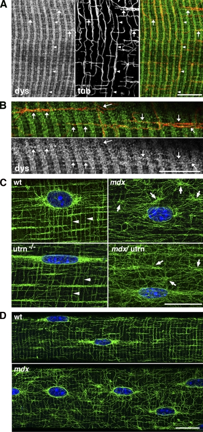
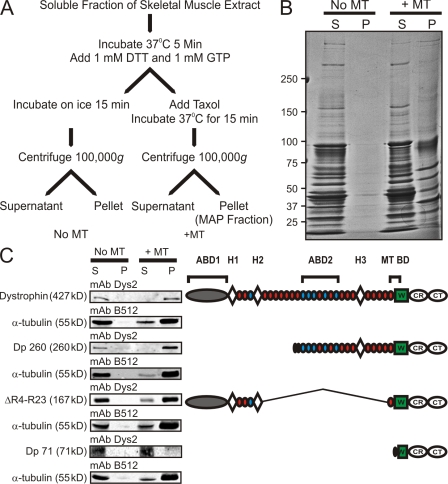
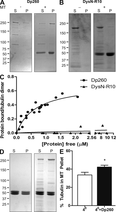
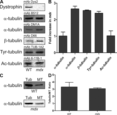
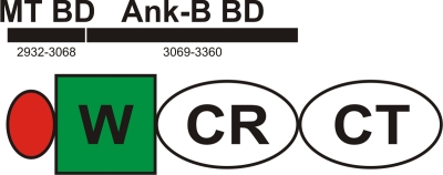
Cytolinkers are giant proteins that can stabilize cells by linking actin filaments, intermediate filaments, and microtubules (MTs) to transmembrane complexes. Dystrophin is functionally similar to cytolinkers, as it links the multiple components of the cellular cytoskeleton to the transmembrane dystroglycan complex. Although no direct link between dystrophin and MTs has been documented, costamere-associated MTs are disrupted when dystrophin is absent. Using tissue-based cosedimentation assays on mice expressing endogenous dystrophin or truncated transgene products, we find that constructs harboring spectrinlike repeat 24 through the first third of the WW domain cosediment with MTs. Purified Dp260, a truncated isoform of dystrophin, bound MTs with a K(d) of 0.66 microM, a stoichiometry of 1 Dp260/1.4 tubulin heterodimer at saturation, and stabilizes MTs from cold-induced depolymerization. Finally, alpha- and beta-tubulin expression is increased approximately 2.5-fold in mdx skeletal muscle without altering the tubulin-MT equilibrium. Collectively, these data suggest dystrophin directly organizes and/or stabilizes costameric MTs and classifies dystrophin as a cytolinker in skeletal muscle.


Figures

Figure 1.
Figure 1.
Dystrophin guides MTs at the surface of the muscle fibers and is necessary for proper MT organization. (A) Isolated muscle fibers from the extensor digitorum longus of 7-wk-old wt mice were costained for dystrophin (left) and α-tubulin (middle). The right panel shows that MTs (red) follow dystrophin (green) bands for long distances both transversely (arrowheads) and longitudinally (arrows). (B) At a higher magnification, dystrophin staining is granular; MTs are studded with dystrophin “dots.” Arrows indicate longitudinal MTs that follow dystrophin. (C) Muscle fibers from the extensor digitorum longus of 7-wk-old wt, mdx, utrn−/−, and mdx/utrn−/− mice were stained with DM1A anti–α-tubulin and Hoechst dye. Both wt and utrn−/− fibers show the lattice of transverse and longitudinal MTs characteristic of fast fibers (arrowheads). In mdx and mdx/utrn−/− fibers, the regularity of the lattice is lost, and mostly oblique MTs originate from cytoplasmic nucleation points (arrows). (D) Peripherally nucleated prenecrotic muscle fibers from 24-d-old mdx mice also displayed MT disorganization, indicating that MT derangement occurred before muscle cell necrosis and regeneration. Bars: (A and B) 10 µm; (C and D) 20 µm.
Figure 2.
Figure 2.
Dystrophin cosediments with MTs in skeletal muscle extracts. (A) Flowchart of tissue MT cosedimentation assay. (B) Coomassie blue–stained SDS-PAGE showing supernatant (S) and pellet (P) fractions in conditions that favored MT depolymerization or polymerization. The pellet fraction in the presence of MTs represents the MAP fraction of skeletal muscle. The molecular mass standards (given in kilodaltons) are indicated on the left. (C, left) Western blot analysis of tissue cosedimentation assay from skeletal muscle extracts of wt mice expressing dystrophin or mdx mice transgenically expressing Dp260, ΔR4-R23, and Dp71. (right) Diagrammatic representation of constructs analyzed in tissue cosedimentation. ABD, actin-binding domain; H, hinge region; W, WW domain; CR, cysteine-rich domain; CT, carboxy-terminal domain; MT BD, MT-binding domain.
Figure 3.
Figure 3.
Dp260 binds and stabilizes MTs in vitro. (A) Coomassie blue–stained SDS-PAGE showing the supernatant (S) and pellet (P) fractions of purified Dp260 in the presence and absence of MTs. Dp260 shifted to the pellet fraction when MTs were present, indicating that Dp260 binds MTs. (B) Coomassie blue–stained SDS-PAGE showing the supernatant and pellet fractions of DysN-R10 in the presence and absence of MTs. The presence of MTs did not cause a shift of DysN-R10 into the pellet fraction, indicating that DysN-R10 does not bind MTs. (C) Concentration-dependent binding of Dp260 to taxol-stabilized MTs (2 µM tubulin) from three independent experiments. DysN-R10 did not show significant MT-binding activity. (D) Coomassie blue–stained SDS-PAGE of the supernatant and pellet fractions of MTs induced to depolymerize by incubating at 4°C in the presence or absence of 1 µM Dp260. (E) Quantification of tubulin in the MT fraction when induced to depolymerize by incubating at 4°C (n = 6). The presence of 1 µM Dp260 significantly (t test; *, P ≤ 0.05) increased the amount of tubulin in the MT fraction, indicating that Dp260 stabilizes MTs. Error bars represent mean ± SEM. (A, B, and D) Molecular mass standards (given in kilodaltons) are indicated on the left.
Figure 4.
Figure 4.
Mdx mice exhibit tubulin misregulation in the absence of an altered tubulin–MT equilibrium. (A) Representative Western blots of tubulin levels in skeletal muscle extracts from wt and mdx mice. (B) Quantification of tubulin levels from three wt and three mdx extracts. (C) Representative Western blots of tubulin (tub) and MT fractions of skeletal muscle extracts from wt and mdx mice using mAb DM1A. (D) Quantification of tubulin–MT equilibrium from five wt and four mdx tibialis anterior muscles. Loss of dystrophin does not affect the tubulin–MT equilibrium. Error bars represent mean ± SEM.
Figure 5.
Figure 5.
Diagrammatic representation of MT- and ankyrin-B–binding domains of dystrophin. Numbers indicate amino acids of full-length dystrophin. H, hinge region; W, WW domain; CR, cysteine-rich domain; CT, carboxy-terminal domain; MT BD, MT-binding domain; Ank-B BD, ankyrin-B–binding domain.

References

    1. Altman A., Szyper-Kravitz M., Shoenfeld Y. 2007. Colchicine-induced rhabdomyolysis.Clin. Rheumatol. 26:2197–2199 - PubMed
    1. Andra K., Lassmann H., Bittner R., Shorny S., Fassler R., Propst F., Wiche G. 1997. Targeted inactivation of plectin reveals essential function in maintaining the integrity of skin, muscle, and heart cytoarchitecture.Genes Dev. 11:3143–3156 - PMC - PubMed
    1. Ayalon G., Davis J.Q., Scotland P.B., Bennett V. 2008. An ankyrin-based mechanism for functional organization of dystrophin and dystroglycan.Cell. 135:1189–1200 - PubMed
    1. Barton E.R., Morris L., Musaro A., Rosenthal N., Sweeney H.L. 2002. Muscle-specific expression of insulin-like growth factor I counters muscle decline in mdx mice.J. Cell Biol. 157:137–148 - PMC - PubMed
    1. Bhosle R.C., Michele D.E., Campbell K.P., Li Z., Robson R.M. 2006. Interactions of intermediate filament protein synemin with dystrophin and utrophin.Biochem. Biophys. Res. Commun. 346:768–777 - PubMed

Publication types