. 2009 Aug 10;186(3):343-53.
doi: 10.1083/jcb.200902146. Epub 2009 Aug 3.

A role of histone H3 lysine 4 methyltransferase components in endosomal trafficking

Affiliations

A role of histone H3 lysine 4 methyltransferase components in endosomal trafficking

Zhuojin Xu et al. J Cell Biol. .

Abstract

Histone lysine methyltransferase complexes are essential for chromatin organization and gene regulation. Whether any of this machinery functions in membrane traffic is unknown. In this study, we report that mammal Dpy-30 (mDpy-30), a subunit of several histone H3 lysine 4 (H3K4) methyltransferase (H3K4MT) complexes, resides in the nucleus and at the trans-Golgi network (TGN). The TGN targeting of mDpy-30 is mediated by BIG1, a TGN-localized guanine nucleotide exchange factor for adenosine diphosphate ribosylation factor GTPases. Altering mDpy-30 levels changes the distribution of cation-independent mannose 6-phosphate receptor (CIMPR) without affecting that of TGN46 or transferrin receptor. Our experiments also indicate that mDpy-30 functions in the endosome to TGN transport of CIMPR and that its knockdown results in the enrichment of internalized CIMPR and recycling endosomes near cell protrusions. Much like mDpy-30 depletion, the knockdown of Ash2L or RbBP5, two other H3K4MT subunits, leads to a similar redistribution of CIMPR. Collectively, these results suggest that mDpy-30 and probably H3K4MT play a role in the endosomal transport of specific cargo proteins.


Figures

Figure 1.
Figure 1.
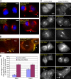
Imaging analyses of perinuclear mDpy-30 in HeLa cells. (A–C) Cells were fixed in 3% formaldehyde, permeabilized with 0.1% saponin, immunostained, and visualized using conventional (A and B) or confocal (C) microscopy. Arrows indicate sites of colocalization. (A) Colocalization analysis of mDpy-30 and recycling endosomes (EGFP-Rab11), late endosomes (EGFP-Rab7), and lysosomes (Lamp1). (B) Colocalization analysis of mDpy-30 and the cis-Golgi network/cis-Golgi (p115), medial-Golgi (GRASP55), or TGN (TGN46) marker. (C) Colocalization analysis of mDpy-30 and p115 or TGN46. A representative image from single z section is shown (the full z stack for both are available in the JCB DataViewer; https://doi.org/10.1083/jcb.200902146.dv). Pearson's correlations were calculated from each image in the z stack of confocal microscopy images of the perinuclear regions (defined by p115 or TGN46) using the colocalization analysis from Image-Pro Plus 3.6 software (Media Cybernetics; Student's t test, P = 5.02393 × 10⁻¹²; n = 14). Error bars indicate the standard deviation of the values obtained from individual cells. Bars, 10 µm.
Figure 2.
Figure 2.
Subcellular distribution analyses of CIMPR, TGN46, and TfnR in HeLa cells depleted of or overexpressing mDpy-30. All siRNAs were used at a concentration of 20 nM, and transfected cells were analyzed 48 h after transfection. Unless noted otherwise, the values shown in all bar graphs and Western blots are the mean results obtained from at least three independent experiments. For imaging analyses, >500 cells were counted in each experiment. (A) Subcellular distributions of CIMPR, TGN46, or TfnR in HeLa cells treated with control nontargeting or mDpy-30 siRNAs. Cells were processed as described in Fig. 1. Arrows indicate cell protrusions. (B) Enrichment of CIMPR near cell protrusions (indicated by arrows) was confirmed by phalloidin F-actin costaining, and the fraction of cells displaying enriched CIMPR near protrusions was scored. (C) Western blot analysis of the CIMPR protein level after the knockdown of mDpy-30. The numbers represent the mean value of six independent experiments. (D) The effect of mDpy-30 overexpression on the localization of CIMPR or β-GalT1 (compare asterisks with arrows). Only those cells exhibiting a high level of exogenous mDpy-30 were counted. The experiment was repeated once, and the mean value is shown. (E) The protein level of CIMPR in cells overexpressing mDpy-30. (B and E) Error bars indicate the standard deviation of the values obtained from independent experiments. Bars, 10 µm.
Figure 3.
Figure 3.
The role of mDpy-30 in the endosome to TGN transport of CIMPR in HeLa cells. Transfection and staining were performed as described in Figs. 2 and 1, respectively, except in G and J where 0.1% Triton X-100 was used to assure even permeabilization of the nuclear membrane among cells. The values shown in all bar graphs and Western blots are the mean results obtained from at least three independent experiments. In the case of imaging analyses, >500 cells were counted in each experiment. Except in J, arrows and the arrowhead indicate cell protrusions. (A) The impact of mDpy-30 knockdown on the protein level of CD8-CIMPR fusion expressed in a HeLa stable line. (B and C) The effect of mDpy-30 depletion on the endosomal transport of CD8-CIMPR monitored at different time points after internalization as described in Materials and methods. Cells were analyzed by eye using the following criteria. In B, cells were scored as perinuclear or mixed perinuclear/peripheral if internalized CD8-CIMPR could be detected as a distinct perinuclear structure in close proximity to TGN46. However, cells lacking such structure were scored as peripheral. In C, cells were scored based on whether internalized CD8-CIMPR became enriched near cell protrusions as described in Fig. 2 B. (D) The influence of knocking down mDpy-30 on the endosomal transport of endogenous CIMPR in HeLa cells was assessed after adding anti-CIMPR antibody to the culture medium for 1 h at 37°C. (E) The comparison of the endosomal transport of CD8-CIMPR and TfnR in mDpy-30–depleted HeLa stable cells. Surface TfnR and CD8-CIMPR were labeled with biotinylated transferrin and the anti-CD8 antibody, respectively, and their endosomal transport was examined at 5, 10, and 20 min after internalization. (F) EEA1 staining was absent at the CD8-CIMPR–enriched cell protrusions. Nu, nucleus. (G and H) The influence of depleting Ash2L or RbBP5 (two H3K4MT components) and SUV39H1 (a component of H3K9MT) on the targeting of internalized CD8-CIMPR to cell protrusions. The effectiveness of Ash2L and RbBP5 siRNAs was examined using immunofluorescence and Western blot (see I) analyses. Cells were scored according to the criteria described in Fig. 2 B, and the result is shown in the bar graph. (I) The impact of knocking down Ash2L or RbBP5 on the protein level of mDpy-30. The level of GAPDH (glyceraldehyde-3-phosphate dehydrogenase) was used as a loading control. (J) The effects of Ash2L or RbBP5 overexpression on the localization of CIMPR or β-GalT1 (compare asterisks with arrows). (B, C, H, and J) Error bars indicate the standard deviation of the values obtained from independent experiments. Bars, 10 µm.
Figure 4.
Figure 4.
Characterization of the CIMPR-enriched compartment at cell protrusions. Transfection and staining were conducted as described in Figs. 2 and 1, respectively. Arrows indicate cell protrusions. (A) The localization of STX10 and VAMP3 after mDpy-30 knockdown. (B) Colocalization analysis of internalized CD8-CIMPR and STX10 or VAMP3 near cell protrusions. Internalization was performed as described in Fig. 3. Nu, nucleus. (C and D) The influence of mDpy-30 knockdown on the localization of various endosomal Rab proteins in HeLa–CD8-CIMPR stable cells. EGFP fusions of Rab proteins were transfected into cells pretreated with a nontargeting control or mDpy-30 siRNA2, and transfected cells were scored according to the criteria described in Fig. 2 B. The numbers in the bar graph represent the mean result from five independent experiments (n > 500 in each experiment). Error bars indicate the standard deviation of the values obtained from independent experiments. Similar results were obtained with mDpy-30 siRNA1, albeit with less effect (not depicted). Bars, 10 µm.
Figure 5.
Figure 5.
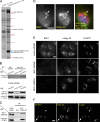
BIG1-mediated TGN targeting of mDpy-30. (A) Identification of mDpy-30–interacting proteins by immunopurification and tandem mass spectroscopy from CV-1 cells stably expressing an EGFP fusion of mDpy-30 (see Materials and methods). Question marks indicate bands that remain unidentified. (B and C) Coimmunoprecipitation of endogenous mDpy-30 and transfected HA-tagged BIG1 (B) or endogenous BIG1 (C). An equal amount of cell lysate was used for coimmunoprecipitation in all samples. IP, immunoprecipitation; WB, Western blot. (D) Colocalization of BIG1 and mDpy-30 (indicated by arrows). (E) Influence of depleting BIG1 on the perinuclear targeting of mDpy-30. The punctate staining of BIG1 in the cytoplasm is background. (F) Impact of overexpressing BIG1 on the perinuclear targeting of mDpy-30 (compare asterisks with arrows). Bars, 10 µm.

References

    1. Cai H., Reinisch K., Ferro-Novick S. 2007. Coats, tethers, Rabs, and SNAREs work together to mediate the intracellular destination of a transport vesicle. Dev. Cell. 12:671–682. - PubMed
    1. Caswell P.T., Norman J.C. 2006. Integrin trafficking and the control of cell migration. Traffic. 7:14–21. - PubMed
    1. Caswell P., Norman J. 2008. Endocytic transport of integrins during cell migration and invasion. Trends Cell Biol. 18:257–263. - PubMed
    1. Chapman H.A. 1997. Plasminogen activators, integrins, and the coordinated regulation of cell adhesion and migration. Curr. Opin. Cell Biol. 9:714–724. - PubMed
    1. Chen J.W., Murphy T.L., Willingham M.C., Pastan I., August J.T. 1985. Identification of two lysosomal membrane glycoproteins. J. Cell Biol. 101:85–95. - PMC - PubMed

Publication types

Substances