. 2009 Aug 18;120(7):553-9.
doi: 10.1161/CIRCULATIONAHA.108.821082. Epub 2009 Aug 3.

Drug-sensitized zebrafish screen identifies multiple genes, including GINS3, as regulators of myocardial repolarization

Affiliations

Drug-sensitized zebrafish screen identifies multiple genes, including GINS3, as regulators of myocardial repolarization

David J Milan et al. Circulation. .

Abstract

Background: Cardiac repolarization, the process by which cardiomyocytes return to their resting potential after each beat, is a highly regulated process that is critical for heart rhythm stability. Perturbations of cardiac repolarization increase the risk for life-threatening arrhythmias and sudden cardiac death. Although genetic studies of familial long-QT syndromes have uncovered several key genes in cardiac repolarization, the major heritable contribution to this trait remains unexplained. Identification of additional genes may lead to a better understanding of the underlying biology, aid in identification of patients at risk for sudden death, and potentially enable new treatments for susceptible individuals.

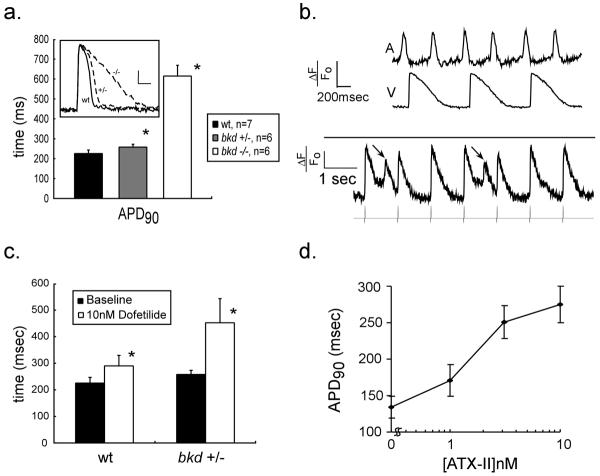
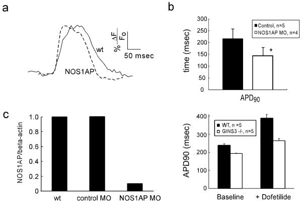
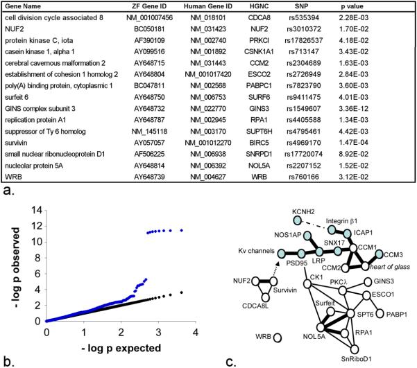
Methods and results: We extended and refined a zebrafish model of cardiac repolarization by using fluorescent reporters of transmembrane potential. We then conducted a drug-sensitized genetic screen in zebrafish, identifying 15 genes, including GINS3, that affect cardiac repolarization. Testing these genes for human relevance in 2 concurrently completed genome-wide association studies revealed that the human GINS3 ortholog is located in the 16q21 locus, which is strongly associated with QT interval.

Conclusions: This sensitized zebrafish screen identified 15 novel myocardial repolarization genes. Among these genes is GINS3, the human ortholog of which is a major locus in 2 concurrent human genome-wide association studies of QT interval. These results reveal a novel network of genes that regulate cardiac repolarization.


Figures

Figure 1
Figure 1. Parallels between zebrafish and human cardiac repolarization
(1a) Ventricular action potential durations (APD) in wildtype (wt) and breakdance heterozygotes (+/−) and homozygotes (−/−) at 6 days post fertilization. * denotes p<0.05. (Inset) Typical ventricular action potentials are displayed for wildtype (wt), breakdance heterozygote (+/−) and homozygote (−/−) embryos. The heterozygote action potential is subtly prolonged, while the homozygote recording shows marked action potential prolongation. Vertical calibration bar denotes 20% ΔF/F0, horizontal bar denotes 100ms. (1b) Upper panel: simultaneous atrial and ventricular voltage recordings from a breakdance (−/−) heart showing the mechanism of 2:1 atrioventricular block: action potentials are so prolonged in the ventricle that alternate atrial impulses encroach on the refractory plateau of the previous ventricular repolarization. Lower panel: Early afterdepolarizations (EADs) (arrows) observed in breakdance (−/−) embryos during ventricular pacing; the pacing train is shown below the action potential recording. EADs appear as spontaneous depolarizations occurring before repolarization is complete and prior to the subsequent paced beat. (1c) Heterozygote breakdance embryos display increased sensitivity to IKr block (10nM dofetilide). (1d) Ventricular action potentials prolong in a dose-dependent fashion with ATX-II. Hearts were paced at 120bpm in ATX-II experiments. All values are expressed as mean +/− S.D.
Figure 2
Figure 2. Genetic Modifiers of Repolarization
(2a) Representative voltage recordings from wild type and NOS1AP morphant hearts are shown. (2b) Action potentials are shortened in NOS1AP morphant hearts compared to mismatch morpholino injected controls. (2c) NOS1AP morpholino injection results in specific knockdown of processed NOS1AP mRNA. qPCR results are displayed in arbitrary units normalized to beta-actin levels. (2d) Ventricular action potentials are shortened in GINS3 homozygous mutants, both at baseline and after treatment with 20nM dofetilide, hearts are paced at 90ppm. All values are expressed as mean +/− S.D (* denotes p<0.05).
Figure 3
Figure 3. Tests of 15 genes discovered in zebrafish reveals role in human QT interval duration
(3a) Table of human orthologs of zebrafish repolarization genes with gene ID number and HGNC name. The SNPs column lists the SNP with strongest evidence for QT interval association in the QTSCD GWAS along with the resulting unadjusted p values. (3b) Quantile-quantile plot of observed versus expected p values. Observed p-values from the QTSCD GWAS, in blue, from tests of all SNPs lying within 150kb of the human orthologs of zebrafish repolarization genes are plotted against p values that would be anticipated by chance alone. Departures from the linear, expected p values (black line) are due to markers with a signal for genetic association. (3c) Interactions between known repolarization genes (blue symbols) and the genes identified in the current study are depicted. Single lines indicate genetic interactions supported by data from multiple model organisms. Bold lines show direct physical interactions. A dashed line represents a physical interaction that may not be direct. The arrow represents a downstream regulatory effect, the mechanism of which is unknown. Supporting data are summarized in Supplemental Table 2.

References

    1. Priori SG, Napolitano C, Schwartz PJ, Grillo M, Bloise R, Ronchetti E, Moncalvo C, Tulipani C, Veia A, Bottelli G, Nastoli J. Association of long QT syndrome loci and cardiac events among patients treated with beta-blockers. Jama. 2004;292:1341–4. - PubMed
    1. Siscovick DS, Raghunathan TE, Rautaharju P, Psaty BM, Cobb LA, Wagner EH. Clinically silent electrocardiographic abnormalities and risk of primary cardiac arrest among hypertensive patients. Circulation. 1996;94:1329–33. - PubMed
    1. Brooksby P, Batin PD, Nolan J, Lindsay SJ, Andrews R, Mullen M, Baig W, Flapan AD, Prescott RJ, Neilson JM, Cowley AJ, Fox KA. The relationship between QT intervals and mortality in ambulant patients with chronic heart failure. The united kingdom heart failure evaluation and assessment of risk trial (UK-HEART) Eur Heart J. 1999;20:1335–41. - PubMed
    1. Roden DM. Drug-induced prolongation of the QT interval. N Engl J Med. 2004;350:1013–22. - PubMed
    1. Wilke RA, Lin DW, Roden DM, Watkins PB, Flockhart D, Zineh I, Giacomini KM, Krauss RM. Identifying genetic risk factors for serious adverse drug reactions: current progress and challenges. Nat Rev Drug Discov. 2007;6:904–16. - PMC - PubMed

Publication types