. 2009 Sep;119(9):2550-63.
doi: 10.1172/JCI33288. Epub 2009 Aug 3.

Increased local expression of coagulation factor X contributes to the fibrotic response in human and murine lung injury

Affiliations

Increased local expression of coagulation factor X contributes to the fibrotic response in human and murine lung injury

Chris J Scotton et al. J Clin Invest. 2009 Sep.

Abstract

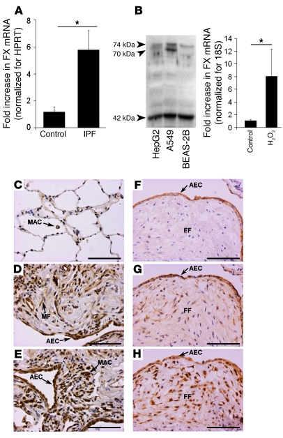
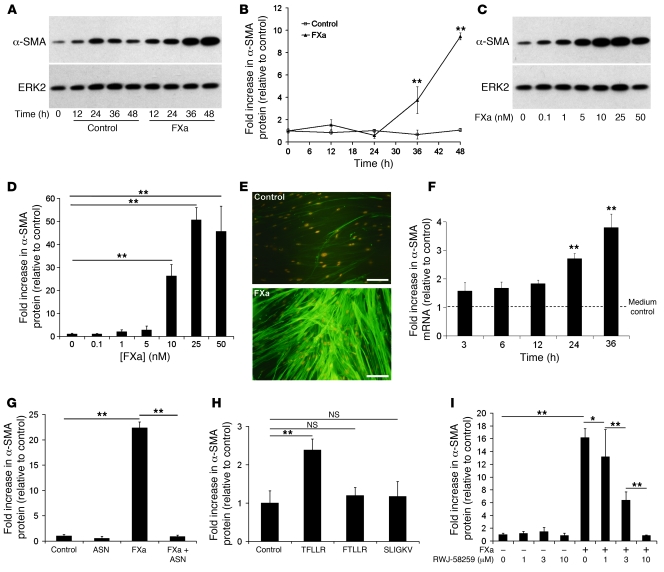
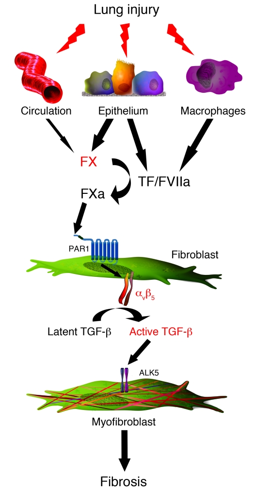
Uncontrolled activation of the coagulation cascade contributes to the pathophysiology of several conditions, including acute and chronic lung diseases. Coagulation zymogens are considered to be largely derived from the circulation and locally activated in response to tissue injury and microvascular leak. Here we report that expression of coagulation factor X (FX) is locally increased in human and murine fibrotic lung tissue, with marked immunostaining associated with bronchial and alveolar epithelia. FXa was a potent inducer of the myofibroblast differentiation program in cultured primary human adult lung fibroblasts via TGF-beta activation that was mediated by proteinase-activated receptor-1 (PAR1) and integrin alphavbeta5. PAR1, alphavbeta5, and alpha-SMA colocalized to fibrotic foci in lung biopsy specimens from individuals with idiopathic pulmonary fibrosis. Moreover, we demonstrated a causal link between FXa and fibrosis development by showing that a direct FXa inhibitor attenuated bleomycin-induced pulmonary fibrosis in mice. These data support what we believe to be a novel pathogenetic mechanism by which FXa, a central proteinase of the coagulation cascade, is locally expressed and drives the fibrotic response to lung injury. These findings herald a shift in our understanding of the origins of excessive procoagulant activity and place PAR1 central to the cross-talk between local procoagulant signaling and tissue remodeling.


Figures

Figure 1
Figure 1. Bleomycin-induced lung injury increases local expression of FX/FXa in the murine lung.
(A) Functional annotation analysis of the genes regulated in mice 14 days after bleomycin instillation, showing the percentage of genes statistically overrepresented within each gene ontology category, arranged in descending order of significance. (B) FX mRNA levels in mouse lung homogenates at 7 and 14 days after saline (Sal; n = 3) or bleomycin (Bleo; n = 5), as assessed by real-time RT-PCR. Data are mean ± SEM. (C) Photomicrographs of representative histological sections showing FX/FXa immunoreactivity in mouse lungs 7 and 14 days after saline or bleomycin (n = 4 per group). Immunostaining increased in bleomycin-instilled mice and localized mainly to alveolar (AEC) and bronchial (BEC) epithelial cells as well as alveolar macrophage (MAC) and myofibroblasts (MF) in fibrotic foci. Inset shows an IgG isotype control section, revealing no apparent staining (original magnification, ×200). Scale bars: 100 μm. (D) Microvascular leak was determined by efflux of intravenously injected EBD into BALF. Data are mean ± SEM (n = 5 per group). (E) Active FXa levels in BALF were determined by chromogenic assay. Data are mean ± SEM (n = 5 per group). *P < 0.05, **P < 0.01, ***P < 0.001, ANOVA.
Figure 2
Figure 2. FX expression is locally upregulated in the human UIP lung.
(A) FX expression in microdissected alveolar septae from human IPF and donor lungs, analyzed by real-time RT-PCR. Data are mean ± SEM (n = 4 patients per group). (B) FX was expressed by HepG2, A549, and BEAS-2B cells in vitro, as demonstrated by Western blot. FX mRNA expression in A549 was upregulated by 1 hour treatment with 400 μM H2O2. Data are mean ± SEM, representative of 3 independent experiments. (CE) Photomicrographs of representative histological sections showing FX/FXa immunolocalization in the lung biopsy specimens of the Brompton cohort. (C) Weak staining was observed in normal control biopsy tissue (n = 6), which mainly localized to alveolar macrophages. (D and E) A marked increase in staining was observed in fibrotic biopsy specimens (n = 7), localized mainly to alveolar epithelial cells, but also to macrophages and myofibroblasts in fibrotic foci. (FH) Photomicrographs of TF (F), FVII/FVIIa (G), and FX/FXa (H) in serial sections, showing colocalization of all 3 to alveolar epithelium overlying fibrotic foci (FF). Fibroblasts within fibrotic foci were also strongly immunoreactive for FX/FXa. Scale bars: 100 μm. *P = 0.005, Student’s t test.
Figure 3
Figure 3. FXa induces fibroblast-to-myofibroblast differentiation via activation of PAR1, but not of PAR2.
(A and B) Time response of FXa-induced α-SMA protein expression in pHALFs. (A) Representative immunoblot for α-SMA and ERK2 loading control. (B) Quantification by densitometry. (C and D) Dose response of FXa-induced α-SMA protein expression. (C) Representative immunoblots. (D) Quantification by densitometry. (E) Immunocytofluorescence demonstrating α-SMA stress fiber formation in pHALFs after FXa exposure for 36 hours. Scale bars: 40 μm. (F) Effect of FXa stimulation on α-SMA mRNA expression in pHALFs, analyzed by real-time RT-PCR. (G) Effect of antistasin (ASN) on FXa-induced α-SMA protein expression, measured by Western blotting. (H) Effect of synthetic peptide agonists for PAR1 (TFLLR-NH2, 200 μM), PAR2 (SLIGKV-NH2, 200 μM) and scrambled peptide control (FTLLR-NH2, 200 μM) on α-SMA protein expression. (I) Effect of the selective PAR1 antagonist RWJ-58259 on FXa-induced α-SMA protein expression. Data are mean ± SD, representative of 3 separate experiments with 3 replicates per experimental group. *P < 0.05, **P < 0.001, ANOVA.
Figure 4
Figure 4. FXa-induced α-SMA protein expression is dependent on TGF-β activity.
(A) Effect of TFLLR-NH2 on TGF-β activation. pHALFs were grown in coculture with tMLECs (see Methods). Activation was blocked by the pan-specific TGF-β–neutralizing antibody 1D11. (B and C) Time course of SMAD2 phosphorylation by FXa. (B) Representative immunoblot for pSMAD2 and total SMAD2/3 loading control. (C) Densitometric analysis. (D and E) Effect of ALK5 inhibition with SB431542 (D) and SD-208 (E) on FXa-induced α-SMA protein expression, analyzed by Western blotting. Cell cultures were preincubated with inhibitors for 30 minutes prior to 36 hours FXa exposure. Densitometric analyses are shown. (F and G) Effect of αvβ5-neutralizing antibodies on FXa-induced α-SMA protein expression. Cell cultures were preincubated with 10 μg/ml anti-αvβ5 or isotype control antibody for 1 hour prior to 36 hours FXa exposure. (F) Representative immunoblots for α-SMA and ERK2 loading control. (G) Densitometric analysis (mean ± SEM). (H) Effect of Y-27632 on FXa-induced α-SMA protein expression. Cell cultures were preincubated with Y-27632 for 1 hour prior to 36 hours FXa exposure; densitometric analysis is shown (mean ± SEM). (I and J) Effect of αvβ5-neutralizing antibodies on FXa-induced SMAD2 phosphorylation. Cell cultures were preincubated with 10 μg/ml anti-αvβ5 or isotype control antibody for 1 hour prior to 10 hours FXa exposure. (I) Representative immunoblots. (J) Densitometric analysis, representative of 2 separate experiments (mean ± SEM). Unless otherwise indicated, data (mean ± SD) are representative of 3 separate experiments, with 3 replicates per group. *P < 0.05, **P < 0.001, ANOVA.
Figure 5
Figure 5. Immunolocalization of α-SMA, αvβ5, and PAR1 in human IPF lung.
(A and B) Photomicrographs of representative 3-μm serial cryostat sections showing immunoreactivity for α-SMA (A) and αvβ5 (B). Spindle-shaped cells within this fibrotic region were positive for both proteins. (C and D) Photomicrographs of representative 3-μm serial paraffin sections showing immunoreactivity for α-SMA (C) and PAR1 (D). Again, spindle-shaped cells within a fibrotic region of IPF lung were positive for both markers. Scale bars: 100 μm.
Figure 6
Figure 6. Inhibition of FXa reduces bleomycin-induced lung fibrosis.
(A and B) Effect of ZK 807834 on FXa-induced α-SMA protein expression in vitro. (A) Representative Western blot. (B) Densitometric analysis. Data are mean ± SEM based on 3 replicates per group. (C) FXa inhibition attenuated lung collagen accumulation after bleomycin instillation in mice, as measured by reverse-phase HPLC quantitation of lung hydroxyproline in acid hydrolysates of pulverized lung. ZK, ZK 807834; veh, vehicle. Data are mean ± SEM increase in lung collagen accumulation relative to the saline plus vehicle group. Values were obtained from groups of 6 (saline plus vehicle and bleomycin plus ZK 807834), 3 (saline plus ZK 807834), or 7 (bleomycin plus vehicle) mice. *P < 0.05, **P < 0.001, ANOVA.
Figure 7
Figure 7. Mechanism underlying the contribution of FX to lung fibrosis.
After lung injury, both locally produced and circulation-derived FX contributes to increased FXa activity in the fibrotic lung. FXa induces its profibrotic effects via activation of PAR1 and subsequent differentiation of fibroblast into the myofibroblast phenotype. This is dependent on the activation of the αvβ5 integrin, leading to the activation of the major profibrotic cytokine TGF-β.

References

    1. Contrino J., Hair G., Kreutzer D.L., Rickles F.R. In situ detection of tissue factor in vascular endothelial cells: correlation with the malignant phenotype of human breast disease. Nat. Med. 1996;2:209–215. doi: 10.1038/nm0296-209. - DOI - PubMed
    1. Matetzky S., et al. Smoking increases tissue factor expression in atherosclerotic plaques: implications for plaque thrombogenicity. Circulation. 2000;102:602–604. - PubMed
    1. Idell S., et al. Procoagulant activity in bronchoalveolar lavage in the adult respiratory distress syndrome. Contribution of tissue factor associated with factor VII. Am. Rev. Respir. Dis. 1987;136:1466–1474. - PubMed
    1. Sapru A., Wiemels J.L., Witte J.S., Ware L.B., Matthay M.A. Acute lung injury and the coagulation pathway: Potential role of gene polymorphisms in the protein C and fibrinolytic pathways. Intensive Care Med. 2006;32:1293–1303. doi: 10.1007/s00134-006-0223-5. - DOI - PubMed
    1. Anthoni C., et al. Tissue factor: a mediator of inflammatory cell recruitment, tissue injury, and thrombus formation in experimental colitis. J. Exp. Med. 2007;204:1595–1601. doi: 10.1084/jem.20062354. - DOI - PMC - PubMed

Publication types

MeSH terms

LinkOut - more resources