. 2009 Aug 4;4(8):e6498.
doi: 10.1371/journal.pone.0006498.

Human mesenchymal stem cells self-renew and differentiate according to a deterministic hierarchy

Affiliations

Human mesenchymal stem cells self-renew and differentiate according to a deterministic hierarchy

Rahul Sarugaser et al. PLoS One. .

Abstract

Background: Mesenchymal progenitor cells (MPCs) have been isolated from a variety of connective tissues, and are commonly called "mesenchymal stem cells" (MSCs). A stem cell is defined as having robust clonal self-renewal and multilineage differentiation potential. Accordingly, the term "MSC" has been criticised, as there is little data demonstrating self-renewal of definitive single-cell-derived (SCD) clonal populations from a mesenchymal cell source.

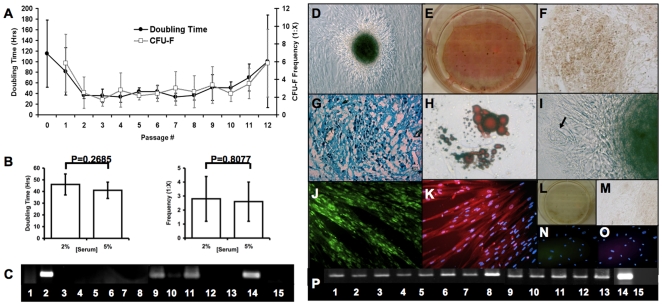
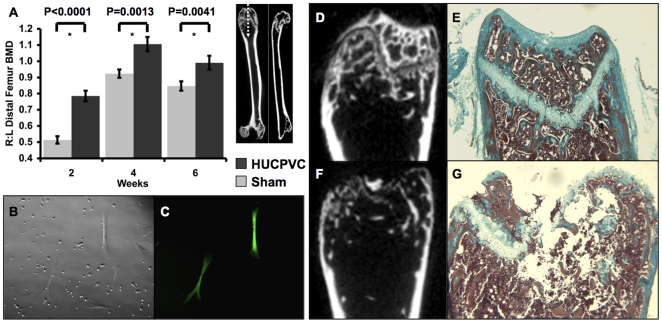
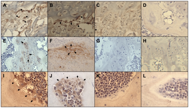
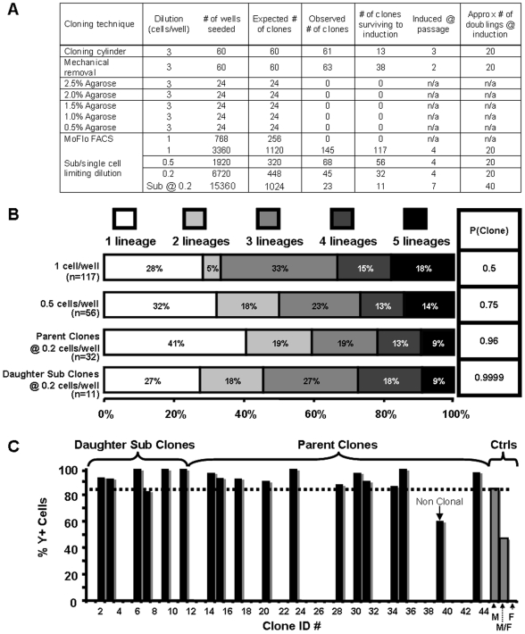
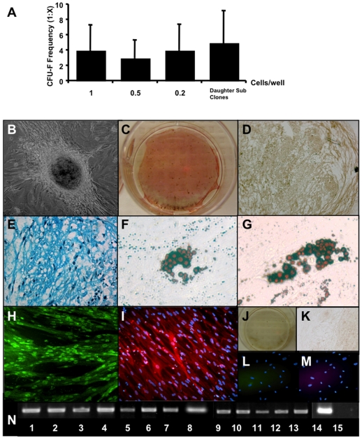
Methodology/principal findings: Here we show that a tractable MPC population, human umbilical cord perivascular cells (HUCPVCs), was capable of multilineage differentiation in vitro and, more importantly, contributed to rapid connective tissue healing in vivo by producing bone, cartilage and fibrous stroma. Furthermore, HUCPVCs exhibit a high clonogenic frequency, allowing us to isolate definitive SCD parent and daughter clones from mixed gender suspensions as determined by Y-chromosome fluorescent in situ hybridization.

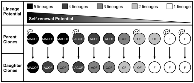
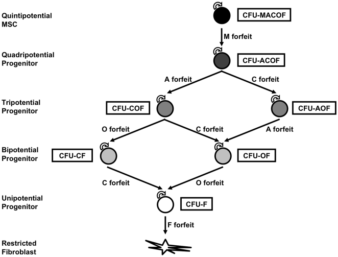
Conclusions/significance: Analysis of the multilineage differentiation capacity of SCD parent clones and daughter clones enabled us to formulate a new hierarchical schema for MSC self-renewal and differentiation in which a self-renewing multipotent MSC gives rise to more restricted self-renewing progenitors that gradually lose differentiation potential until a state of complete restriction to the fibroblast is reached.


Conflict of interest statement

Competing Interests: The authors have declared that no competing interests exist.

Figures

Figure 1
Figure 1. HUCPVC populations exhibit rapid proliferation, high clonogenicity and multipotential capacity in vitro.
HUCPVC populations maintained stable proliferation and clonogenic frequency from passage 2 through 9 (A, n = 11 different cords), with no significant difference in either parameter at passage 5 (B, n = 7 different cords). Data are represented as mean +/− s.d. In standard culture conditions, HUCPVCs expressed Collagen IA1, Desmin and MyoD [lanes 2, 9 and 11 respectively, all other lanes as represented in (P)] as determined by RT PCR (C). HUCPVCs could further differentiate into bone, cartilage, adipose and muscle in vitro. With induction, bone nodules were observed in culture (D) that stained with Von Kossa (black), and were surrounded by alkaline phosphatase-high expressing cells (E). Cartilage pellet cultures of HUCPVCs expressed collagen II (F) and glycosaminoglycans that stained with Alcian blue (G). HUCPVC-derived adipocytes stained with Oil Red O (H) and occasionally formed spontaneously (arrow) in association with bone nodules (I). Myogenically-induced HUCVPCs expressed high levels of MyoD (J) and fast skeletal myosin light chain (FSMLC) (K) in multinucleated HUCPVC myotubes. Negative controls are non-induced cells stained with Von Kossa and alkaline phosphatase (L), and secondary-only antibody staining for collagen II (M), MyoD (N) and FSMLC (O) stained cultures. (Field widths: D,F = 628 µm; E,L = 3.5 mm; G,J,K,N,O = 315 µm; H,I = 86 µm). RT PCR analysis (P) demonstrated upregulation of the following lineage-specific genes: Runx2 (1), collagen IA1 (2), osteopontin (3), osteocalcin (4), Sox9 (5), collagen II (6), lipoprotein lipase (LPL) (7), aggregan (8), MyoD (9), Myf5 (10), desmin (11), myosin heavy chain (MHC) (12), FSMLC (13), GAPDH (14) and RT- control (15). (Cells from 9 cords, some of which are common to other assays, were employed in these functional phenotypic and gene expression data).
Figure 2
Figure 2. HUCPVCs survive and regenerate damaged mesenchymal tissues in vivo.
HUCPVCs were synthetically active and survived at least six weeks in vivo when implanted into the intrafemoral space of NOD-scid mice as observed by the presence of GFP-labeled cells among mouse cells flushed from the marrow into culture (B,C). When the distal ends of the femurs were analyzed by micro-computed tomography (µCT), it was determined that these cells produced significantly more bone mineral density (BMD) than sham controls at 2,4 and 6 weeks (A, values are means±s.d.). µCT analysis and Masson's Trichrome stained longitudinal sections of injected femurs showed that HUCPVCs induced significantly more repair of bone and cartilage at 2 weeks (D,E) compared to sham controls (F,G). HUCPVC populations from a total 6 different cords were employed to generate this data and that shown in Figure 3.
Figure 3
Figure 3. HUCPVCs display multipotential capacity in vivo.
When labeled with HuNu (human nuclear antigen), human cells (arrows) were observed in the growth plate of the distal femurs (A,B field widths = 140 µm). These cells were associated with the presence of human-specific collagen II (arrows) in the extracellular matrix surrounding individual chondrocytes (E,F field widths = 140 µm and 212 µm respectively). Human-specific osteocalcin was also observed on the osteoid of newly forming bone. Arrows indicate human-specific osteocalcin was present surrounding individual cells that produced a collagen-like matrix on the surface of the bone (I,J field widths = 110 µm). No staining was observed in sham-injected femurs or negative controls for HuNu (C,D), collagen II (G,H) or osteocalcin (K,L) respectively (same field widths as experimentals).
Figure 4
Figure 4. Clonally pure HUCPVC populations can be isolated by rigorous cell seeding.
Five different methods were used in an attempt to generate definitive single-cell-derived (SCD) clones from a total of 12 cords, some of which were employed in other assays, (A). Clonal colonies generated by seeding mixed male and female suspensions of HUCPVCs at 1, 0.5, and 0.2 cells/well, the latter of which were sub-cloned (daughters) at 0.2 cells/well, displayed increasing probabilities of SCD isolation along with differential multipotential capacities (B). When analyzed by fluorescent in situ hybridization (FISH) with a Y-chromosome-specific probe, all the daughters (clones 1–11) as well as all but one (clone 39) of the parent clones (clones 12–44) seeded at 0.2 cells/well were found to be single gender derived, and accordingly determined to be SCD clones (C).
Figure 5
Figure 5. Clonally pure HUCPVC populations display multipotent capacity in vitro.
CFU-F frequencies of HUCPVCs derived by seeding at 1, 0.5, and 0.2 cells/well, the latter of which were sub-cloned (daughters) at 0.2 cells/well, were not found to be significantly different from each other (A). Clone 11 (B-N) demonstrated identical capacity to its parent (clone 35), maintaining the ability to differentiate into all five lineages assayed. Under induction, bone nodules were observed in culture (B) that stained with Von Kossa (black), and were surrounded by alkaline phosphatase-high expressing cells (C). Cartilage pellet cultures of HUCPVCs expressed collagen II (D) and glycosaminoglycans that stained with Alcian blue (E). HUCPVC-derived adipocytes stained with Oil Red O (F,G). Myogenically-induced HUCVPCs expressed high levels of MyoD (H) and FSMLC (I) in multinucleated HUCPVC myotubes. Negative controls were uninduced cells stained with Von Kossa and alkaline phosphatase (J), and secondary-only antibody staining for collagen II (K), MyoD (L) and FSMLC (M) stained cultures. (Field widths: B,D,K = 628 µm; C,J = 3.5 mm; E,H,I,L,M = 315 µm; F,G = 86 µm). RT-PCR analysis (O) demonstrated upregulation of the following lineage-specific genes: Runx2 (1), collagen IA1 (2), osteopontin (3), osteocalcin (4), Sox9 (5), collagen II (6), LPL (7), aggregan (8), MyoD (9), Myf5 (10), desmin (11), MHC (12), FSMLC (13), GAPDH (14) and RT negative control (15). (MACOF represents a clone that was able to differentiate into all 5 lineages assayed). HUCPVC harvests from 27 different cords were employed to generate this data.
Figure 6
Figure 6. Clonally pure HUCPVC populations display differential self-renewal capacity in vitro.
When the differentiation capacities of the 11 daughter clones were compared to those of their parents, it was found that they either maintained equipotency (self-renewing clones) or forfeited the ability to differentiate into one or more lineages (non-self-renewing clones). (Self-renewing clones are represented with a semi-circular arrow and non-self-renewing clones without a semi-circular arrow).
Figure 7
Figure 7. Hierarchy of MSC differentiation.

References

    1. Friedenstein AJ, Gorskaja JF, Kulagina NN. Fibroblast precursors in normal and irradiated mouse hematopoietic organs. Exp Hematol. 1976;4:267–274. - PubMed
    1. Castro-Malaspina H, Gay RE, Resnick G, Kapoor N, Meyers P, et al. Characterization of human bone marrow fibroblast colony-forming cells (CFU-F) and their progeny. Blood. 1980;56:289–301. - PubMed
    1. Kuznetsov SA, Friedenstein AJ, Robey PG. Factors required for bone marrow stromal fibroblast colony formation in vitro. Br J Haematol. 1997;97:561–570. - PubMed
    1. Kuznetsov SA, Krebsbach PH, Satomura K, Kerr J, Riminucci M, et al. Single-colony derived strains of human marrow stromal fibroblasts form bone after transplantation in vivo. J Bone Miner Res. 1997;12:1335–1347. - PubMed
    1. Zuk PA, Zhu M, Ashjian P, De Ugarte DA, Huang JI, et al. Human adipose tissue is a source of multipotent stem cells. Molecular Biology of the Cell. 2002;13:4279–4295. - PMC - PubMed

Publication types