. 2009 Aug 4:9:268.
doi: 10.1186/1471-2407-9-268.

Alterations of BCCIP, a BRCA2 interacting protein, in astrocytomas

Affiliations

Alterations of BCCIP, a BRCA2 interacting protein, in astrocytomas

Jingmei Liu et al. BMC Cancer. .

Abstract

Background: Loss of heterozygosity of chromosome 10q26 has been shown to be associated with the aggressiveness of astrocytic tumors (or astrocytomas), but the responsible gene(s) residing in this region has not been fully identified. The BCCIP gene is located at chromosome 10q26. It encodes a BRCA2 and CDKN1A (p21) interacting protein. Previous studies have shown that down-regulation of BCCIP impairs recombinational DNA repair, G1/S cell cycle checkpoint, p53 trans-activation activity, cytokinesis, and chromosome stability, suggesting a potential role of BCCIP in cancer etiology. In this study, we investigated whether BCCIP is altered in astrocytomas.

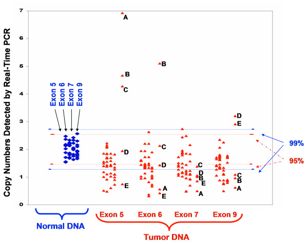
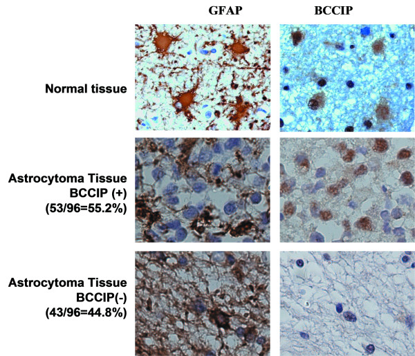
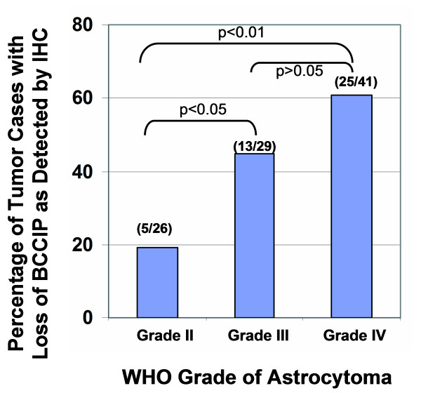
Methods: Genomic DNA from 45 cases of grade IV astrocytic tumor (glioblastoma) tissues and 12 cases of normal tissues were analyzed by quantitative PCR. The BCCIP protein expression in 96 cases of grade II-IV astrocytic tumors was detected by immunohistochemistry (IHC). IHC staining of glial fibrillary acid protein (GFAP), a marker for astrocytic cells, was used to identify cells of the astrocytic lineage.

Results: We found that BCCIP protein is expressed in normal cells with positive staining of GFAP. However, BCCIP protein expression was not detectable in approximately 45% of all astrocytic tumors, and in > 60% in the grade IV glioblastoma. About 45% glioblastoma have significant (p < 0.01) reduction of BCCIP gene copy number when compared to normal DNA. Furthermore, the frequency of lacking BCCIP expression is associated with the aggressiveness of astrocytic tumors.

Conclusion: Our data implicate a role of BCCIP in astrocytic tumorigenesis, and lack of BCCIP may be used as a marker for astrocytomas.


Figures

Figure 1
Figure 1
Copy numbers of BCCIP exons detected by quantitative Real-Time PCR. The copy number of exons 5, 6, 7, and 9 were measured in 10 cases of normal brain tissue DNA (diamond marker), and 45 cases of glioblastoma DNA (see Materials and Methods for details). The 95% and 99% confidence ranges of the copy number in normal tissues are marked. Many tumor DNAs showed reduction of BCCIP copy numbers. However, five cases of tumor DNA (marked as cases A-E) have reduced BCCIP copy numbers at some exons but increased in others. These re-arrangements likely cause inactivation of the BCCIP gene.
Figure 2
Figure 2
Lack of BCCIP expression in brain tumors. Serial tissue sections were stained for GFAP and BCCIP separately. Both GFAP and BCCIP are stained in brown color. Hematoxylin (blue) was used to counter stain the nuclei. Shown are example brain tissues stained with antibodies against GFAP and BCCIP (magnification is 40 × 10). Top panel is a representative non-tumor section. The middle panel is an example section of BCCIP positive tumor, and the bottom panel shows a BCCIP negative tumor section.
Figure 3
Figure 3
Frequency of loss of BCCIP protein expression in different WHO grades of astrocytomas [30]. Shown is the percentage of astrocytomas that are BCCIP negative. The p-values indicate the statistic values between the indicated tumor grade groups.

References

    1. Rasheed BK, Fuller GN, Friedman AH, Bigner DD, Bigner SH. Loss of heterozygosity for 10q loci in human gliomas. Genes Chromosomes Cancer. 1992;5:75–82. doi: 10.1002/gcc.2870050111. - DOI - PubMed
    1. Karlbom AE, James CD, Boethius J, Cavenee WK, Collins VP, Nordenskjold M, Larsson C. Loss of heterozygosity in malignant gliomas involves at least three distinct regions on chromosome 10. Hum Genet. 1993;92:169–74. doi: 10.1007/BF00219686. - DOI - PubMed
    1. Balesaria S, Brock C, Bower M, Clark J, Nicholson SK, Lewis P, de Sanctis S, Evans H, Peterson D, Mendoza N. Loss of chromosome 10 is an independent prognostic factor in high-grade gliomas. Br J Cancer. 1999;81:1371–7. doi: 10.1038/sj.bjc.6693403. - DOI - PMC - PubMed
    1. Steck PA, Lin H, Langford LA, Jasser SA, Koul D, Yung WK, Pershouse MA. Functional and molecular analyses of 10q deletions in human gliomas. Genes Chromosomes Cancer. 1999;24:135–43. doi: 10.1002/(SICI)1098-2264(199902)24:2<135::AID-GCC6>3.0.CO;2-A. - DOI - PubMed
    1. Fujisawa H, Reis RM, Nakamura M, Colella S, Yonekawa Y, Kleihues P, Ohgaki H. Loss of heterozygosity on chromosome 10 is more extensive in primary (de novo) than in secondary glioblastomas. Lab Invest. 2000;80:65–72. doi: 10.1038/labinvest.3780009. - DOI - PubMed

Publication types