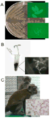
Bacillus anthracis physiology and genetics
- PMID: 19654018
- PMCID: PMC2784286
- DOI: 10.1016/j.mam.2009.07.004
Bacillus anthracis physiology and genetics
Abstract
Bacillus anthracis is a member of the Bacillus cereus group species (also known as the "group 1 bacilli"), a collection of Gram-positive spore-forming soil bacteria that are non-fastidious facultative anaerobes with very similar growth characteristics and natural genetic exchange systems. Despite their close physiology and genetics, the B. cereus group species exhibit certain species-specific phenotypes, some of which are related to pathogenicity. B. anthracis is the etiologic agent of anthrax. Vegetative cells of B. anthracis produce anthrax toxin proteins and a poly-d-glutamic acid capsule during infection of mammalian hosts and when cultured in conditions considered to mimic the host environment. The genes associated with toxin and capsule synthesis are located on the B. anthracis plasmids, pXO1 and pXO2, respectively. Although plasmid content is considered a defining feature of the species, pXO1- and pXO2-like plasmids have been identified in strains that more closely resemble other members of the B. cereus group. The developmental nature of B. anthracis and its pathogenic (mammalian host) and environmental (soil) lifestyles of make it an interesting model for study of niche-specific bacterial gene expression and physiology.
Figures


References
-
- Ackermann HW, Roy R, Martin M, Murthy MR, Smirnoff WA. Partial characterization of a cubic Bacillus phage. Can J Microbiol. 1978;24:986–993. - PubMed
-
- Ash C, Collins MD. Comparative analysis of 23S ribosomal RNA gene sequences of Bacillus anthracis and emetic Bacillus cereus determined by PCR-direct sequencing. FEMS Microbiol Lett. 1992;73:75–80. - PubMed
Publication types
MeSH terms
Substances
Grants and funding
LinkOut - more resources
Full Text Sources

