

Advanced correlative light/electron microscopy: current methods and new developments using Tokuyasu cryosections
- PMID: 19654103
- PMCID: PMC2778083
- DOI: 10.1369/jhc.2009.954214
Advanced correlative light/electron microscopy: current methods and new developments using Tokuyasu cryosections
Abstract
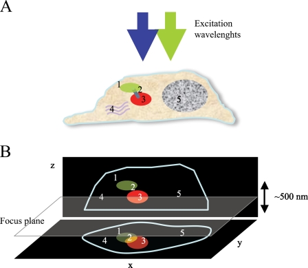
Microscopy is an essential tool for analysis of cellular structures and function. With the advent of new fluorescent probes and super-resolution light microscopy techniques, the study of dynamic processes in living cells has been greatly facilitated. Fluorescence light microscopy provides analytical, quantitative, and three-dimensional (3D) data with emphasis on analysis of live cells using fluorescent markers. Sample preparation is easy and relatively inexpensive, and the use of appropriate tags provides the ability to track specific proteins of interest. Of course, only electron microscopy (EM) achieves the highest definition in terms of ultrastructure and protein labeling. To fill the gap between light microscopy and EM, correlative light and electron microscopy (CLEM) strategies have been developed. In particular, hybrid techniques based upon immuno-EM provide sensitive protein detection combined with high-resolution information on cell structures and protein localization. By adding the third dimension to EM with electron tomography (ET) combined with rapid freezing, CLEM techniques now provide additional tools for quantitative 3D analysis. Here, we overview the major methods applied and highlight the latest advances in the field of CLEM. We then focus on two selected techniques that use cryosections as substrate for combined biomolecular imaging. Finally, we provide a perspective of future developments in the field.
Figures


Similar articles
-
High data output method for 3-D correlative light-electron microscopy using ultrathin cryosections.Methods Mol Biol. 2013;950:417-37. doi: 10.1007/978-1-62703-137-0_23. Methods Mol Biol. 2013. PMID: 23086888
-
3D HDO-CLEM: cellular compartment analysis by correlative light-electron microscopy on cryosection.Methods Cell Biol. 2012;111:95-115. doi: 10.1016/B978-0-12-416026-2.00006-6. Methods Cell Biol. 2012. PMID: 22857925
-
A workflow for 3D-CLEM investigating liver tissue.J Microsc. 2021 Mar;281(3):231-242. doi: 10.1111/jmi.12967. Epub 2020 Oct 27. J Microsc. 2021. PMID: 33034376
-
Live correlative light-electron microscopy to observe molecular dynamics in high resolution.Microscopy (Oxf). 2016 Aug;65(4):296-308. doi: 10.1093/jmicro/dfw024. Epub 2016 Jul 6. Microscopy (Oxf). 2016. PMID: 27385786 Review.
-
Sugar and ice: Immunoelectron microscopy using cryosections according to the Tokuyasu method.Tissue Cell. 2019 Apr;57:90-102. doi: 10.1016/j.tice.2018.08.010. Epub 2018 Sep 3. Tissue Cell. 2019. PMID: 30201442 Review.
Cited by
-
Serial ultrathin sections to identify ultrastructural localization of GLUT1 molecules in vesicles in brain endothelial cells-correlative light and electron microscopy in depth.Microscopy (Oxf). 2022 Apr 1;71(2):124-131. doi: 10.1093/jmicro/dfac005. Microscopy (Oxf). 2022. PMID: 35157050 Free PMC article.
-
Pyrene Excimer-Based Fluorescent Labeling of Cysteines Brought into Close Proximity by Protein Dynamics: ASEM-Induced Thiol-Ene Click Reaction for High Spatial Resolution CLEM.Int J Mol Sci. 2020 Oct 13;21(20):7550. doi: 10.3390/ijms21207550. Int J Mol Sci. 2020. PMID: 33066147 Free PMC article.
-
FluoroNanogold: an important probe for correlative microscopy.J Chem Biol. 2015 Aug 25;8(4):129-42. doi: 10.1007/s12154-015-0145-1. eCollection 2015 Oct. J Chem Biol. 2015. PMID: 26884817 Free PMC article. Review.
-
Quantitative assessment of specificity in immunoelectron microscopy.J Histochem Cytochem. 2010 Oct;58(10):917-27. doi: 10.1369/jhc.2010.956243. Epub 2010 May 10. J Histochem Cytochem. 2010. PMID: 20458060 Free PMC article.
-
Visualization of Neutrophil Extracellular Traps and Fibrin Meshwork in Human Fibrinopurulent Inflammatory Lesions: III. Correlative Light and Electron Microscopic Study.Acta Histochem Cytochem. 2016 Nov 1;49(5):141-147. doi: 10.1267/ahc.16028. Epub 2016 Oct 26. Acta Histochem Cytochem. 2016. PMID: 27917008 Free PMC article.
References
-
- Abbe E (1873) Beitrage zur Theorie des Mikroskops und der Mikroskopischen Wahrnehmung. Arch Mikr Anat 9:413–468
-
- Agronskaia A, Valentijn J, Van Driel L, Schneijdenberg C, Humbel B, van Bergen en Henegouwen P, Verkleij A, et al. (2008) Integrated fluorescence and trasmission electron microscopy. J Struct Biol 164:183–189 - PubMed
-
- Betzig E, Patterson GH, Sougrat R, Lindwasser OW, Olenych S, Bonifacino JS, Davidson MW, et al. (2006) Imaging intracellular fluorescent proteins at nanometer resolution. Science 313:1642–1645 - PubMed
Publication types
MeSH terms
Substances
LinkOut - more resources
Full Text Sources
Miscellaneous
