. 2009 Oct 2;284(40):27315-26.
doi: 10.1074/jbc.M109.041400. Epub 2009 Aug 4.

A novel protein associated with membrane-type 1 matrix metalloproteinase binds p27(kip1) and regulates RhoA activation, actin remodeling, and matrigel invasion

Affiliations

A novel protein associated with membrane-type 1 matrix metalloproteinase binds p27(kip1) and regulates RhoA activation, actin remodeling, and matrigel invasion

Daisuke Hoshino et al. J Biol Chem. .

Abstract

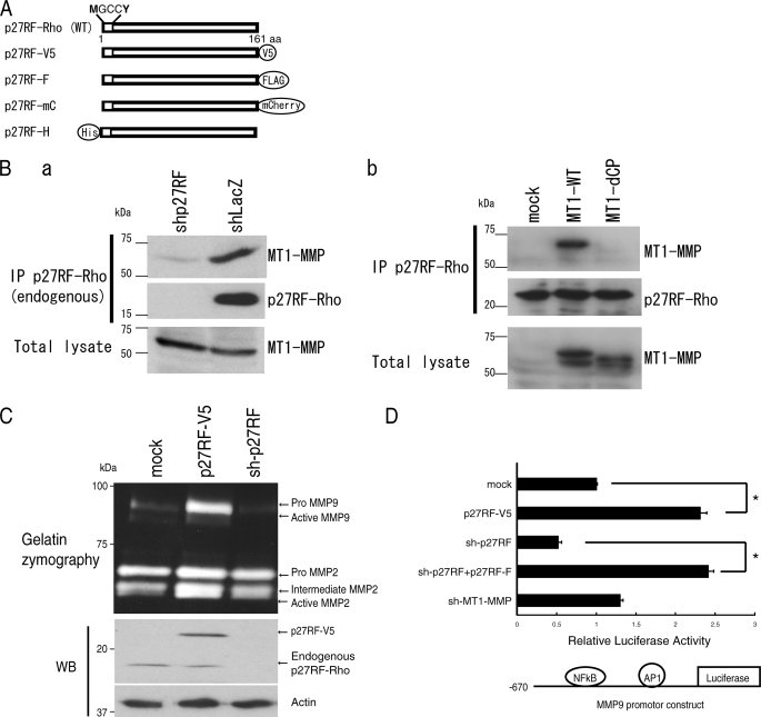
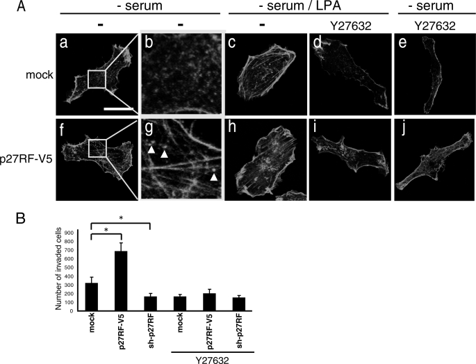
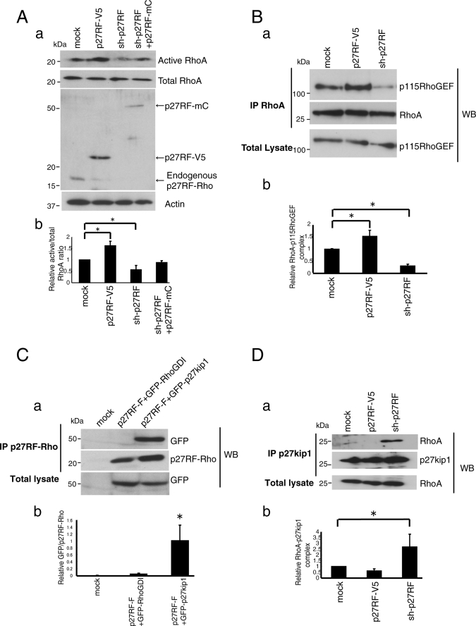
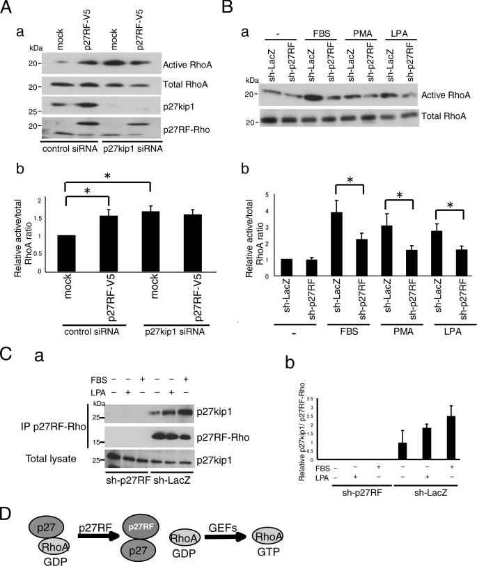
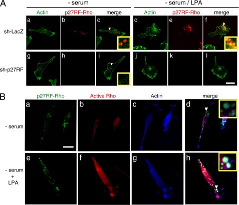
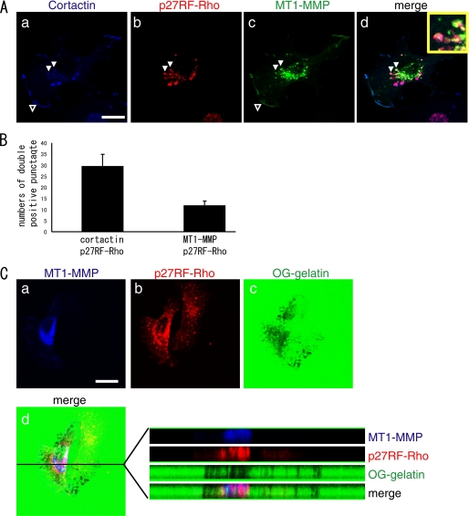
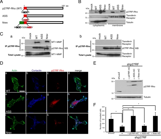
Pericellular proteolysis by membrane-type 1 matrix metalloproteinase (MT1-MMP) plays a pivotal role in tumor cell invasion. Localization of MT1-MMP at the invasion front of cells, e.g. on lamellipodia and invadopodia, has to be regulated in coordination with reorganization of the actin cytoskeleton. However, little is known about how such invasion-related actin structures are regulated at the sites where MT1-MMP localizes. During analysis of MT1-MMP-associated proteins, we identified a heretofore uncharacterized protein. This protein, which we call p27RF-Rho, enhances activation of RhoA by releasing it from inhibition by p27(kip1) and thereby regulates actin structures. p27(kip1) is a well known cell cycle regulator in the nucleus. In contrast, cytoplasmic p27(kip1) has been demonstrated to bind GDP-RhoA and inhibit GDP-GTP exchange mediated by guanine nucleotide exchange factors. p27RF-Rho binds p27(kip1) and prevents p27(kip1) from binding to RhoA, thereby freeing the latter for activation. Knockdown of p27RF-Rho expression renders cells resistant to RhoA activation stimuli, whereas overexpression of p27RF-Rho sensitizes cells to such stimulation. p27RF-Rho exhibits a punctate distribution in invasive human tumor cell lines. Stimulation of the cells with lysophosphatidic acid induces activation of RhoA and induces the formation of punctate actin structures within foci of p27RF-Rho localization. Some of the punctate actin structures co-localize with MT1-MMP and cortactin. Down-regulation of p27RF-Rho prevents both redistribution of actin into the punctate structures and tumor cell invasion. Thus, p27RF-Rho is a new potential target for cancer therapy development.


Figures

FIGURE 1.
FIGURE 1.
The MT1-MMP-associating protein, p27RF-Rho, regulates expression of MMP-9. A, schematic representation of p27RF-Rho and its tagged protein derivatives. p27RF-Rho has consensus sequences for myristoylation and palmitoylation at the N terminus as indicated. p27RF-V5, V5-tagged p27RF-Rho; p27RF-F, FLAG-tagged protein; p27RF-mC, mCherry-tagged protein including a truncation of the 3′-non-coding region of the p27RF mRNA so as to eliminate the sequence targeted by the shRNA for the knockdown experiments; p27RF-H, His-tagged protein. B, a, immunoprecipitation (IP) of endogenous p27RF-Rho by an anti-p27RF-Rho antibody co-precipitated endogenous MT1-MMP as detected by Western blot analysis of the immunoprecipitates using an anti-MT1-MMP antibody. b, p27RF-Rho-FLAG was expressed together with myc-MT1-MMP in HT1080 cells. p27RF-Rho was immunoprecipitated using an anti-FLAG antibody. Proteins bound to the antibody were eluted with FLAG peptides and subjected to Western blot analysis using an anti-Myc antibody. p27RF-Rho precipitated MT1-MMP but not a mutant MT1-MMP lacking the cytoplasmic tail. C, effect of p27RF-Rho on production of gelatinases was analyzed by gelatin zymography. Expression of p27RF-Rho in HT1080 cells was modulated by transfecting an expression plasmid for p27RF-V5, or endogenous protein expression was down-regulated using shRNA (sh-p27RF), respectively. Gelatinase activities of MMP-2 and MMP-9 secreted into the culture media were analyzed by gelatin zymography (upper panel). p27RF-Rho expression was analyzed by Western blot analysis (WB; bottom panels). D, the effect of p27RF-Rho on the mmp-9 promoter was monitored using a luciferase reporter assay in HT 1080. The reporter construct is illustrated at the bottom. Data are shown as the mean ± S.D., n = 5. *, p < 0.05.
FIGURE 2.
FIGURE 2.
p27RF-Rho modulates cell morphology via RhoA activity. A, HT1080 cells stably expressing p27RF-V5 and mock cells (empty vector) were seeded onto collagen coated glass coverslips and treated with LPA and/or Y27632. Actin was visualized with Alexa488-phalloidin. Confocal images were performed using a 63× objective lens (scale bar, 20 μm). B, invasion of Matrigel was analyzed using the indicated HT1080 cell transfectants in the presence or absence of Y27632. The number of cells that had invaded through the Matrigel layer was counted. Data are shown as the mean ± S.D. (n = 3). *, p < 0.05 (Student's t test).
FIGURE 3.
FIGURE 3.
p27RF-Rho promotes the association of GEFs with RhoA. A, GTP-loaded Rho proteins in HT1080 cell lysate were pulled down using GST-Rhotekin for RhoA. RhoA proteins were analyzed by Western blot (WB) using specific antibodies (a, upper panel). Quantification of active RhoA normalized to total RhoA (b, n = 3). *, p < 0.05 (Student's t test). Endogenous and exogenous p27RF-Rho proteins were detected with an anti-p27RF-Rho polyclonal antibody (a, middle panel). B, a, the effect of p27RF-Rho on the association of Myc-RhoA with V5-p115RhoGEF was tested. Myc-RhoA was immunoprecipitated (IP) and the associated GEFs were detected by Western blot analysis. b, quantification of Fig. 2B, a (n = 3). *, p < 0.05 (Student's t test). C, a, GFP-RhoGDI or GFP-p27kip1 was expressed together with p27RF-F in HT1080 cells. p27RF-F was immunoprecipitated using an anti-FLAG antibody, and associated GFP-fused proteins were detected by Western blot assay using an anti-GFP antibody. A representative result of three experiments is indicated. b, quantification of C, a (n = 3). *, p < 0.05 (Student's t test). D, a, Myc-RhoA and FLAG-p27kip1 were expressed in HT1080 cells expressing either p27RF-V5 or sh-p27RF. FLAG-p27kip1 was immunoprecipitated, and Myc-RhoA was detected in the precipitates by Western blot analysis. b, quantification of Fig. 2D, a (n = 3). *, p < 0.05 (Student's t test).
FIGURE 4.
FIGURE 4.
p27RF-Rho activates RhoA via p27kip1 pathway. Aa, effect of p27RF-Rho on RhoA activation in the presence or absence of p27kip1. The expression of p27kip1 in HT1080 cells was knocked down using siRNA. b, quantification of the ratio of active RhoA to total RhoA (n = 3). *, p < 0.05 (Student's t test). B, a, HT1080 cells were serum-starved and treated with FBS, phorbol 12-myristate 13-acetate (PMA), or LPA in the presence or absence of sh-p27RF, and active RhoA was detected. b, quantification of the Western blots (n = 3). *, p < 0.05 (Student's t test). C, a, HT1080 cells were serum-starved and treated with LPA or FBS, then p27kip1 immunoprecipitated (IP) using anti-p27RF-Rho antibody. sh-p27RF was a negative control. b, quantification of C, a (n = 3). D, schematic illustration of the role of p27RF-Rho in the activation of RhoA. p27 kip1 binds GDP-RhoA and prevents GDP-RhoA from binding GEFs. p27RF-Rho binds p27 kip1 and sequesters the latter from GDP-RhoA. GDP-RhoA free from p27 kip1 can be activated by associating with GEFs.
FIGURE 5.
FIGURE 5.
Co-localization of p27RF-Rho, active RhoA, and actin. A, HT1080 cells expressing sh-p27RF or sh-LacZ were seeded onto collagen-coated glass coverslips. Cells were serum-starved and treated with LPA. Actin was visualized with Alexa488-phalloidin (green), and p27RF-Rho was visualized with anti-p27RF-Rho (red). The area indicated by the white arrowhead is magnified in the inset. Confocal images were performed using a 63× objective lens (scale bar, 20 μm). B, after fixation the cells were incubated with soluble GST-RBD to detect active RhoA. Bound GST-RBD was visualized by immunostaining using anti-GST antibody (red). The same samples were double-stained for p27RF-Rho (green) and actin (blue). The area indicated by the white arrowhead is magnified in the inset. CCD images were performed using a 60× objective lens (scale bar, 20 μm).
FIGURE 6.
FIGURE 6.
p27RF-Rho co-localized with MT1-MMP. A, MT1-MMP with a C-terminal Venus-tag was expressed in HT1080. The cells were stained for MT1-MMP (green), p27RF-Rho (red), and cortactin (blue). The area indicated by the white arrowhead is magnified in the inset. Confocal images used a 63× objective lens (scale bar, 20 μm). B, the number of punctate p27RF-Rho signals or MT1-MMP-p27RF-Rho double positive punctate signals were counted. Data are the mean ± S.D. (n = 3). C, a–d, HT1080 cells cultured on Oregon Green-labeled gelatin-coated glass coverslips. After fixation, the cells were stained for p27RF-Rho (red) and MT1-MMP (blue). Note that the degradation of gelatin is specific to the cells expressing exogenous MT1-MMP. d, X-Y and X-Z sections of the gelatin layer and merged image. Confocal images were performed using a 63× objective lens (scale bar, 20 μm).
FIGURE 7.
FIGURE 7.
Membrane targeting signals of p27RF-Rho are necessary to localize to the punctate actin structures and bind to MT1-MMP. A, N-terminal sequences of p27RF-Rho, and its mutants are presented. Gly and Cys indicated by black bold letters correspond to hypothetical myristoylation and palmitoylation sites, respectively. ASS, acylation defective mutant; Nrev, additional acylation sites were fused to the N terminus of the ASS mutant. B, the following proteins were expressed in HT1080 cells; mock (empty vector), wild type (WT), ASS, and Nrev. After cells were lysed and separated into cytoplasmic and membrane fractions, p27RF-Rho was detected by Western blot analysis. Transferrin receptor and tubulin are representative makers for membrane and cytoplasmic proteins, respectively. A representative result of three experiments is indicated. C, Myc-tagged-MT1-MMP (a) and hemagglutinin-tagged transferrin receptor (b) were expressed together with p27RF-F or its mutants in HT1080 cells. The cells were lysed, and p27RF-F was immunoprecipitated (IP) using an anti-FLAG antibody. p27RF-Rho and its mutants bound to the antibody were eluted with FLAG peptides and subjected to Western blot (WB) analysis using anti-Myc or anti-HA antibodies. A representative result of three experiments is indicated. D, p27RF-Rho and its mutant proteins fused to an mCherry tag were expressed in HT1080 cells, and the cells were seeded onto collagen-coated glass coverslips, then serum-starved and treated with LPA. After fixation, the cells were stained for mCherry (red), cortactin (blue), and actin (green). Confocal images were performed using a 63× objective lens (scale bar, 20 μm). E and F, effect of mutant p27RF-Rho on Matrigel invasion. Expression of endogenous p27RF-Rho was knocked down using shRNA in HT1080 cells, and mutant p27RF-Rho proteins were expressed. Proteins were analyzed by Western blot analysis (E). The invasive activity of the cells was analyzed using a Transwell Chamber equipped with a Matrigel-coated filter, and the number of invaded cells was counted. Data are presented as the mean ± S.D. (n = 3). *, p < 0.05 (Student's t test).

References

    1. Egeblad M., Werb Z. (2002) Nat. Rev. Cancer 2, 161–174 - PubMed
    1. Itoh Y., Seiki M. (2006) J. Cell. Physiol. 206, 1–8 - PubMed
    1. Overall C. M., Dean R. A. (2006) Cancer Metastasis Rev. 25, 69–75 - PubMed
    1. Mori H., Tomari T., Koshikawa N., Kajita M., Itoh Y., Sato H., Tojo H., Yana I., Seiki M. (2002) EMBO J. 21, 3949–3959 - PMC - PubMed
    1. Weaver A. M. (2008) Curr. Biol. 18, R362–R364 - PubMed

Publication types

MeSH terms

LinkOut - more resources