

Endogenous HIV-1 Vpr-mediated apoptosis and proteome alteration of human T-cell leukemia virus-1 transformed C8166 cells
- PMID: 19655254
- PMCID: PMC4033303
- DOI: 10.1007/s10495-009-0380-4
Endogenous HIV-1 Vpr-mediated apoptosis and proteome alteration of human T-cell leukemia virus-1 transformed C8166 cells
Abstract
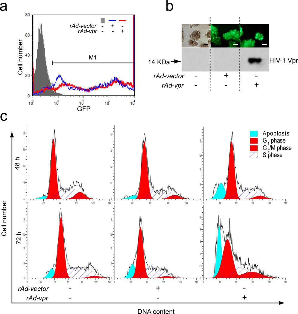
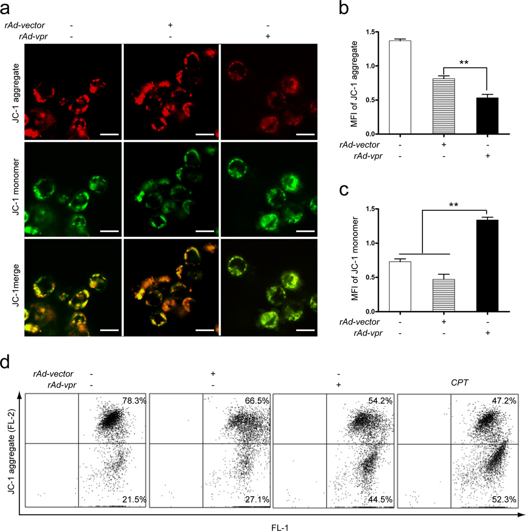
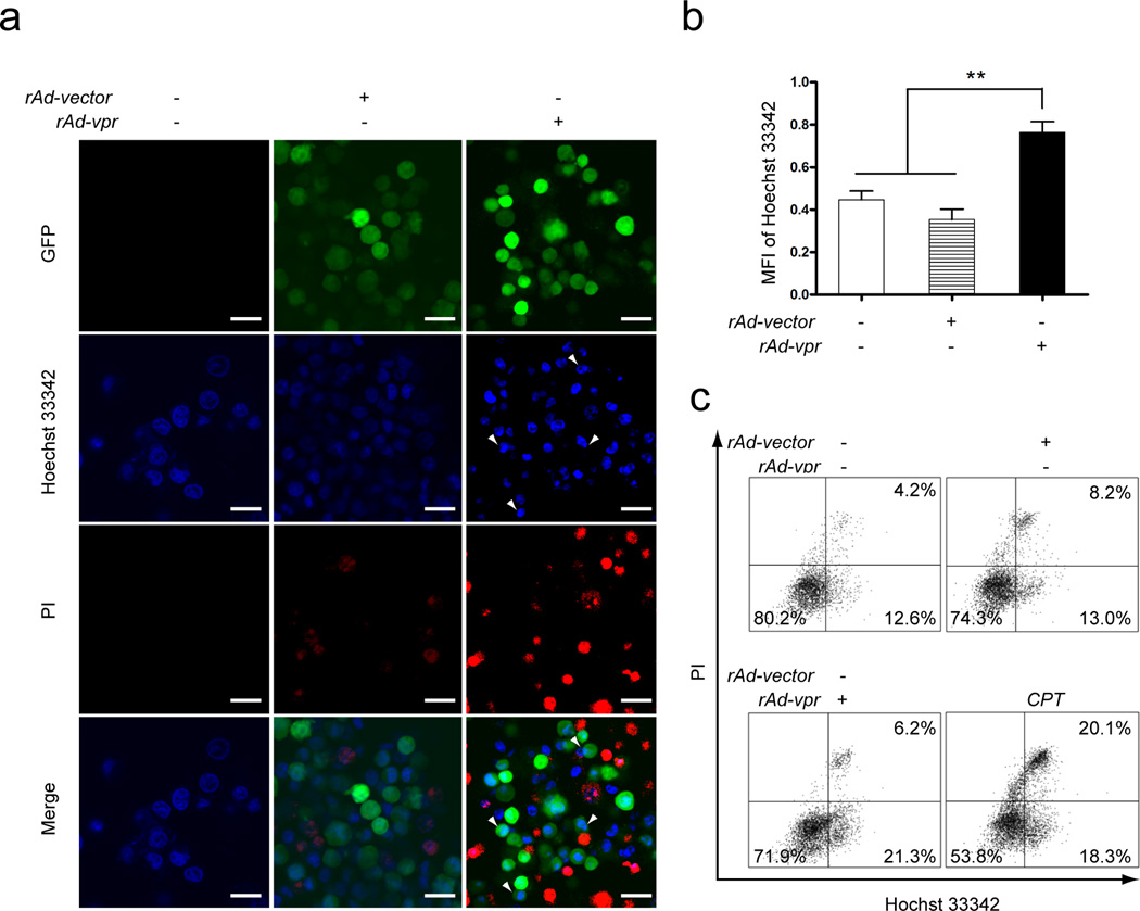
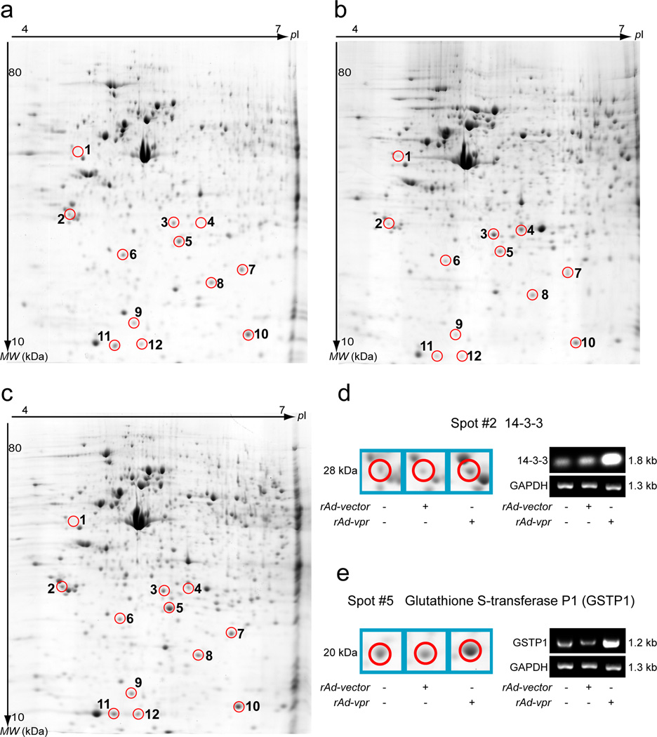
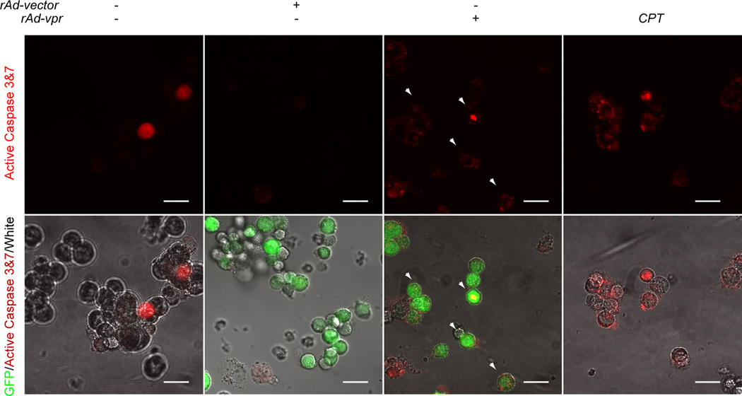
HIV-1 viral protein R (Vpr) can induce cell cycle arrest and cell death, and may be beneficial in cancer therapy to suppress malignantly proliferative cell types, such as adult T-cell leukemia (ATL) cells. In this study, we examined the feasibility of employing the HIV-vpr gene, via targeted gene transfer, as a potential new therapy to kill ATL cells. We infected C8166 cells with a recombinant adenovirus carrying both vpr and GFP genes (rAd-vpr), as well as the vector control virus (rAd-vector). G(2)/M phase cell cycle arrest was observed in the rAd-vpr infected cells. Typical characteristics of apoptosis were detected in rAd-vpr infected cells, including sub-diploid peak exhibition in DNA content assay, the Hoechst 33342 accumulation, apoptotic body formation, mitochondrial membrane potential and plasma membrane integrity loss. The proteomic assay revealed apoptosis related protein changes, exhibiting the regulation of caspase-3 activity indicator proteins (vimentin and Rho GDP-dissociation inhibitor 2), mitochondrial protein (prohibitin) and other regulatory proteins. In addition, the up-regulation of anti-inflammatory redox protein, thioredoxin, was identified in the rAd-vpr infected group. Further supporting these findings, the increase of caspase 3&7 activity in the rAd-vpr infected group was observed. In conclusion, endogenous Vpr is able to kill HTLV-1 transformed C8166 cells, and may avoid the risks of inducing severe inflammatory responses through apoptosis-inducing and anti-inflammatory activities.
Figures
Similar articles
-
The apoptosis-inducing effects of HIV Vpr recombinant eukaryotic expression vectors with different mutation sites on transfected Hela cells.Curr HIV Res. 2009 Sep;7(5):519-25. doi: 10.2174/157016209789346291. Curr HIV Res. 2009. PMID: 19925402
-
Human immunodeficiency virus type 1 vpr induces apoptosis through caspase activation.J Virol. 2000 Apr;74(7):3105-11. doi: 10.1128/jvi.74.7.3105-3111.2000. J Virol. 2000. PMID: 10708425 Free PMC article.
-
Human immunodeficiency virus type 1 Vpr induces cell cycle arrest at the G(1) phase and apoptosis via disruption of mitochondrial function in rodent cells.Microbes Infect. 2006 Mar;8(3):670-9. doi: 10.1016/j.micinf.2005.09.002. Epub 2006 Jan 11. Microbes Infect. 2006. PMID: 16480911
-
HIV1-viral protein R (Vpr) mutations: associated phenotypes and relevance for clinical pathologies.Rev Med Virol. 2016 Sep;26(5):314-29. doi: 10.1002/rmv.1889. Epub 2016 Jun 6. Rev Med Virol. 2016. PMID: 27264019 Review.
-
Partner molecules of accessory protein Vpr of the human immunodeficiency virus type 1.DNA Cell Biol. 2004 Apr;23(4):193-205. doi: 10.1089/104454904773819789. DNA Cell Biol. 2004. PMID: 15142377 Review.
Cited by
-
FTY720 mediates activation suppression and G(0)/G (1) cell cycle arrest in a concanavalin A-induced mouse lymphocyte pan-activation model.Inflamm Res. 2012 Jun;61(6):623-34. doi: 10.1007/s00011-012-0454-6. Epub 2012 Mar 10. Inflamm Res. 2012. PMID: 22407397
-
Host proteome research in HIV infection.Genomics Proteomics Bioinformatics. 2010 Mar;8(1):1-9. doi: 10.1016/S1672-0229(10)60001-0. Genomics Proteomics Bioinformatics. 2010. PMID: 20451157 Free PMC article. Review.
-
Proteomic analysis of PBMCs: characterization of potential HIV-associated proteins.Proteome Sci. 2010 Mar 12;8:12. doi: 10.1186/1477-5956-8-12. Proteome Sci. 2010. PMID: 20222986 Free PMC article.
-
Alterations in the nuclear proteome of HIV-1 infected T-cells.Virology. 2014 Nov;468-470:409-420. doi: 10.1016/j.virol.2014.08.029. Epub 2014 Sep 19. Virology. 2014. PMID: 25240327 Free PMC article.
-
Prohibitin Interacts with envelope proteins of white spot syndrome virus and prevents infection in the red swamp crayfish, Procambarus clarkii.J Virol. 2013 Dec;87(23):12756-65. doi: 10.1128/JVI.02198-13. Epub 2013 Sep 18. J Virol. 2013. PMID: 24049173 Free PMC article.
References
-
- Lilienbaum A, Paulin D. Activation of the human vimentin gene by the Tax human T-cell leukemia virus. I. Mechanisms of regulation by the NF-kappa B transcription factor. J Biol Chem. 1993;268:2180–2188. - PubMed
-
- Miyake K, Inokuchi K, Miyake N, Dan K, Shimada T. HIV vector-mediated targeted suicide gene therapy for adult T-cell leukemia. Gene Ther. 2007;14:1662–1667. - PubMed
-
- Tsukasaki K, Tanosaki S, DeVos S, et al. Identifying progression-associated genes in adult T-cell leukemia/lymphoma by using oligonucleotide microarrays. Int J Cancer. 2004;109:875–881. - PubMed
-
- Kami M, Hamaki T, Miyakoshi S, et al. Allogeneic haematopoietic stem cell transplantation for the treatment of adult T-cell leukaemia/lymphoma. Br J Haematol. 2003;120:304–309. - PubMed
-
- Michelfelder S, Lee MK, deLima-Hahn E, et al. Vectors selected from adeno-associated viral display peptide libraries for leukemia cell-targeted cytotoxic gene therapy. Exp Hematol. 2007;35:1766–1776. - PubMed
Publication types
MeSH terms
Substances
Grants and funding
LinkOut - more resources
Full Text Sources
Research Materials
