

Specificity of the horizontal cell-photoreceptor connections in the zebrafish (Danio rerio) retina
- PMID: 19655401
- PMCID: PMC2841349
- DOI: 10.1002/cne.22135
Specificity of the horizontal cell-photoreceptor connections in the zebrafish (Danio rerio) retina
Abstract
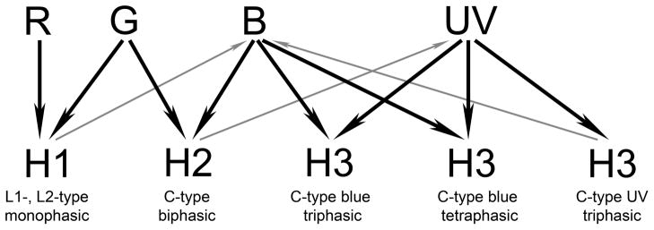
Horizontal cells (HCs) are involved in establishing the center-surround receptive field organization of photoreceptor and bipolar cells. In many species, HCs respond differentially to colors and may play a role in color vision. An earlier study from our laboratory suggested that four types of HCs exist in the zebrafish retina: three cone HCs (H1, H2 and H3) and one rod HC. In this study, we describe their photoreceptor connections. Cones are arranged in a mosaic in which rows of alternating blue (B)- and ultraviolet (UV)-sensitive single cones alternate with rows of red (R)- and green (G)-sensitive double cones; the G cones are adjacent to UV cones and B cones adjacent to R cones. Two small-field (H1 and H2) and two large-field (H3 and rod HC) cells were observed. The cone HC dendritic terminals connected to cones with single boutons, doublets, or rosettes, whereas the rod HCs connected to rods with single boutons. The single boutons/doublets/rosettes of cone HCs were arranged in double rows separated by single rows for H1 cells, in pairs and singles for H2 cells, and in a rectilinear pattern for H3 cells. These connectivity patterns suggest that H1 cells contact R, G, and B cones, H2 cells G, B, and UV cones, and H3 cells B and UV cones. These predictions were confirmed by applying the DiI method to SWS1-GFP retinas whose UV cones express green fluorescent protein. Each rod HC was adjacent to the soma or axon of a DiI-labeled cone HC and connected to 50-200 rods.
Figures


Similar articles
-
Specific connectivity between photoreceptors and horizontal cells in the zebrafish retina.J Neurophysiol. 2016 Dec 1;116(6):2799-2814. doi: 10.1152/jn.00449.2016. Epub 2016 Oct 5. J Neurophysiol. 2016. PMID: 27707811 Free PMC article.
-
Electrical coupling, receptive fields, and relative rod/cone inputs of horizontal cells in the tiger salamander retina.J Comp Neurol. 2006 Nov 20;499(3):422-31. doi: 10.1002/cne.21117. J Comp Neurol. 2006. PMID: 16998920
-
Bipolar cell-photoreceptor connectivity in the zebrafish (Danio rerio) retina.J Comp Neurol. 2012 Nov 1;520(16):3786-802. doi: 10.1002/cne.23168. J Comp Neurol. 2012. PMID: 22907678 Free PMC article.
-
Photoreceptor classes and transmission at the photoreceptor synapse in the retina of the clawed frog, Xenopus laevis.Microsc Res Tech. 2000 Sep 1;50(5):338-46. doi: 10.1002/1097-0029(20000901)50:5<338::AID-JEMT3>3.0.CO;2-I. Microsc Res Tech. 2000. PMID: 10941170 Review.
-
Bipolar Cell Pathways in the Vertebrate Retina.2007 May 24 [updated 2012 Jan 20]. In: Kolb H, Fernandez E, Jones B, Nelson R, editors. Webvision: The Organization of the Retina and Visual System [Internet]. Salt Lake City (UT): University of Utah Health Sciences Center; 1995–. 2007 May 24 [updated 2012 Jan 20]. In: Kolb H, Fernandez E, Jones B, Nelson R, editors. Webvision: The Organization of the Retina and Visual System [Internet]. Salt Lake City (UT): University of Utah Health Sciences Center; 1995–. PMID: 21413382 Free Books & Documents. Review.
Cited by
-
Transmission from the dominant input shapes the stereotypic ratio of photoreceptor inputs onto horizontal cells.Nat Commun. 2014 May 15;5:3699. doi: 10.1038/ncomms4699. Nat Commun. 2014. PMID: 24832361 Free PMC article.
-
Fovea-like Photoreceptor Specializations Underlie Single UV Cone Driven Prey-Capture Behavior in Zebrafish.Neuron. 2020 Jul 22;107(2):320-337.e6. doi: 10.1016/j.neuron.2020.04.021. Epub 2020 May 29. Neuron. 2020. PMID: 32473094 Free PMC article.
-
Diverse Cell Types, Circuits, and Mechanisms for Color Vision in the Vertebrate Retina.Physiol Rev. 2019 Jul 1;99(3):1527-1573. doi: 10.1152/physrev.00027.2018. Physiol Rev. 2019. PMID: 31140374 Free PMC article. Review.
-
Color Processing in Zebrafish Retina.Front Cell Neurosci. 2018 Oct 3;12:327. doi: 10.3389/fncel.2018.00327. eCollection 2018. Front Cell Neurosci. 2018. PMID: 30337857 Free PMC article. Review.
-
Transcription factors underlying photoreceptor diversity.Elife. 2023 Feb 6;12:e81579. doi: 10.7554/eLife.81579. Elife. 2023. PMID: 36745553 Free PMC article.
References
-
- Ahnelt P, Kolb H. Horizontal cells and cone photoreceptors in primate retina: a Golgi-light microscopic study of spectral connectivity. J Comp Neurol. 1994;343(3):387–405. - PubMed
-
- Ammermüller J, Kolb H. Functional architecture of the turtle retina. Progress in Retinal and Eye Research. 1996;15(2):393–433.
-
- Boycott BB. Horizontal cells of mammalian retinae. Neurosci Res Suppl. 1988;8:S97–111. - PubMed
-
- Connaughton VP, Graham D, Nelson R. Identification and morphological classification of horizontal, bipolar, and amacrine cells within the zebrafish retina. J Comp Neurol. 2004;477(4):371–385. - PubMed
Publication types
MeSH terms
Grants and funding
LinkOut - more resources
Full Text Sources
Molecular Biology Databases
Miscellaneous
