. 2009 Oct;9(10):2303-11.
doi: 10.1111/j.1600-6143.2009.02777.x. Epub 2009 Jul 28.

Isolated CD39 expression on CD4+ T cells denotes both regulatory and memory populations

Affiliations

Isolated CD39 expression on CD4+ T cells denotes both regulatory and memory populations

Q Zhou et al. Am J Transplant. 2009 Oct.

Abstract

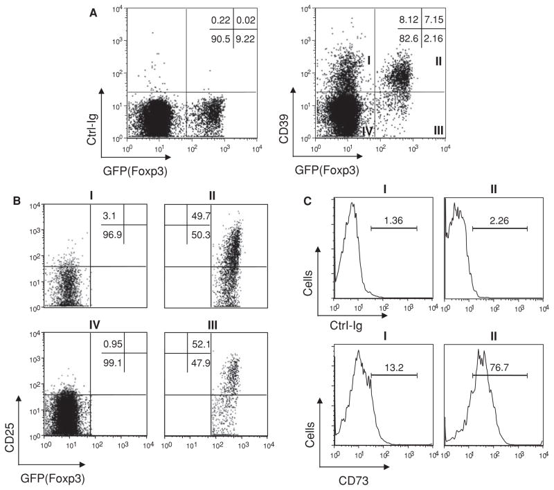
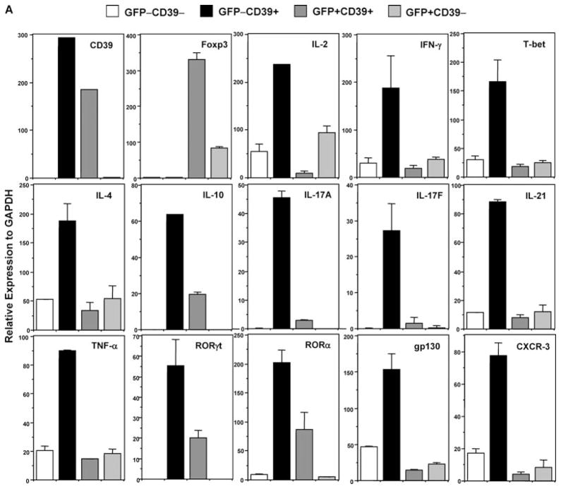
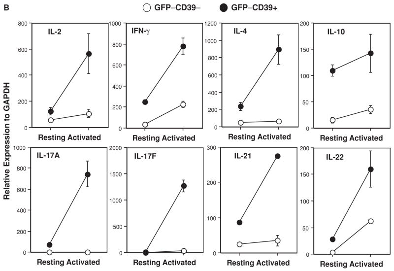
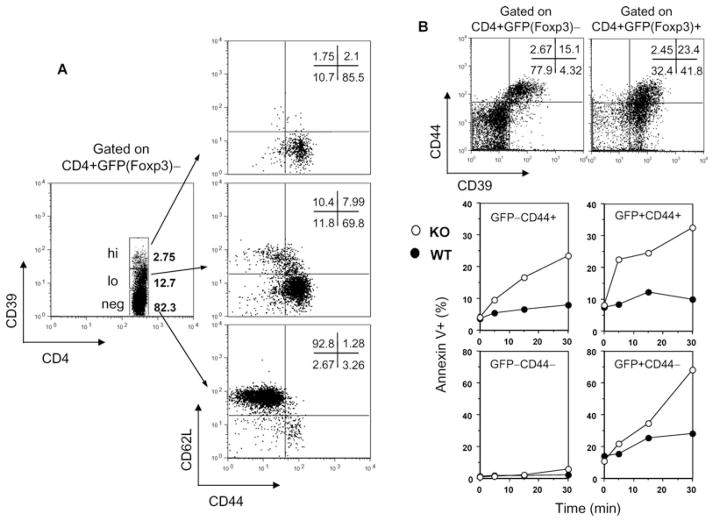
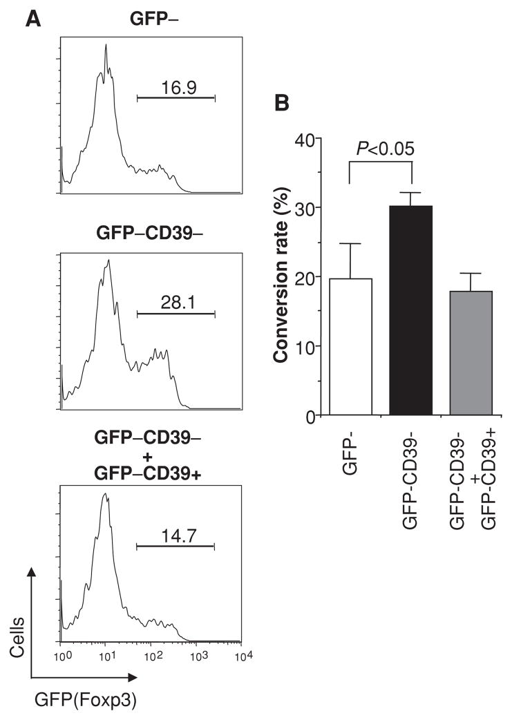
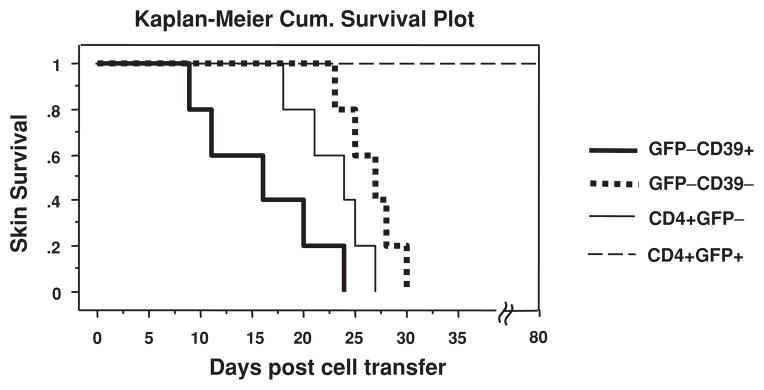
Foxp3(+) regulatory T cells (Tregs) express both ectoenzymes CD39 and CD73, which in tandem hydrolyze pericellular ATP into adenosine, an immunoinhibitory molecule that contributes to Treg suppressive function. Using Foxp3GFP knockin mice, we noted that the mouse CD4(+)CD39(+) T-cell pool contains two roughly equal size Foxp3(+) and Foxp3(-) populations. While Foxp3(+)CD39(+) cells are CD73(bright) and are the bone fide Tregs, Foxp3(-)CD39(+) cells do not have suppressive activity and are CD44(+)CD62L(-)CD25(-)CD73(dim/-), exhibiting memory cell phenotype. Functionally, CD39 expression on memory and Treg cells confers protection against ATP-induced apoptosis. Compared with Foxp3(-)CD39(-) naïve T cells, Foxp3(-)CD39(+) cells freshly isolated from non-immunized mice express at rest significantly higher levels of mRNA for T-helper lineage-specific cytokines IFN-gamma (Th1), IL-4/IL-10 (Th2), IL-17A/F (Th17), as well as pro-inflammatory cytokines, and rapidly secrete these cytokines upon stimulation. Moreover, the presence of Foxp3(-)CD39(+) cells inhibits TGF-beta induction of Foxp3 in Foxp3(-)CD39(-) cells. Furthermore, when transferred in vivo, Foxp3(-)CD39(+) cells rejected MHC-mismatched skin allografts in a much faster tempo than Foxp3(-)CD39(-) cells. Thus, besides Tregs, CD39 is also expressed on pre-existing memory T cells of Th1-, Th2- and Th17-types with heightened alloreactivity.


Conflict of interest statement

Conflict of Interest

The authors have declared that no conflict of interest exists.

Figures

Figure 1
Figure 1. CD39 is expressed on CD4+Foxp3 cells that exhibit memory phenotype
Spleen cells from 6- to 8-week-old naïve Foxp3GFP knockin mice were stained with a rabbit anti-mouse CD39 polyclonal antibody, and live CD4+ T cells were gated for multi-color FACS analysis. (A) Compared with control Ig staining (left panel), anti-CD39 stained positive for subpopulations of cells in Foxp3+ and Foxp3 gates (right panel). (B) Fractions I–IV of (A) were further analyzed for CD25 expression. About 50% of Foxp3+ cells (fractions II and III) are CD25+. Almost all the Foxp3CD39+ cells (fraction I) are CD25. (C) Foxp3+CD39+ cells are CD73bright, whereas Foxp3CD39+ cells are CD73dim/−. Histograms of control Ig (upper panel) or anti-mCD73 (lower panel) staining of fractions I and II were plotted. Data represent more than three independent experiments.
Figure 2
Figure 2. CD4+Foxp3CD39+ cells rapidly produce Th1-, Th2- and Th17-type cytokines
(A) Live CD4+ T cells from unimmunized 6- to 8-week old Foxp3GFP knockin mice were FACS-sorted into fractions I, II, III and IV based on the GFP(Foxp3) versus CD39 staining profile depicted in Figure 1A (right panel). Each sorted fraction was immediately lysed and mRNA extracted for real-time PCR analysis of multiple transcripts. Data represent more than three independent experiments. (B) Rapid production of cytokines by GFPCD39+ Tmem cells. FACS-sorted naïve (GFPCD39) and memory (GFPCD39+) T cells were activated by anti-CD3 and anti-CD28. At 48 hours, aliquots of activated naïve (GFPCD39, empty circle) and memory (GFPCD39+, filled circle) T cells were analyzed by real-time PCR for cytokine messages. Data represent two independent experiments.
Figure 3
Figure 3. CD39 is also a memory T-cell marker and protects against ATP-induced apoptosis
(A) Spleen cells from unimmunized 6- to 8-week-old Foxp3GFP knockin mice were stained and live CD4+Foxp3 T cells were gated into CD39hi, CD39lo and CD39neg populations for analysis of CD44 and CD62L expression. The Foxp3CD39hi population is TEM cells (CD44+CD62L), and the Foxp3CD39lo population contains 70% TEM cells and 8% TCM cells (CD44+CD62L+), whereas the Foxp3CD39neg population is mainly Tnaive cells (CD44CD62L+). (B) CD39 confers protection against ATP-induced apoptosis to CD4+GFPCD44+ Tmem and CD4+GFP+ Treg cells. CD39 knockout mice were bred with Foxp3GFP knockin mice. Freshly isolated splenocytes from WT (filled circle) and CD39 knockout mice (open circle) were incubated with 30 μM ATP for various times at 37°C. Apoptosis of each gated population (upper panel) was measured by Annexin V staining (lower panel). Similar findings were noted when ATP was added at 100 μM. Data represent two independent experiments.
Figure 4
Figure 4. CD4+Foxp3CD39+ cells inhibit the conversion of CD4+Foxp3CD39 cells into iTregs
(A) FACS-sorted CD4+GFP cells (containing CD39+ and CD39 fractions I and IV as in Figure 1A) (upper panel) and CD4+GFPCD39 (fraction IV) (middle panel) were stimulated with anti-CD3 and anti-CD28 in the presence of TGF-β for 3 days. In parallel, sorted CD4+GFPCD39+ cells were added back to the CD4+GFPCD39 culture at the physiological ratio determined during FACS sorting (lower panel). (B) Bar graph with standard deviations from three different experiments.
Figure 5
Figure 5. CD4+Foxp3CD39+ cells reject MHC-mismatched skin grafts at a significantly faster tempo than CD4+Foxp3CD39 cells
FACS-sorted GFPCD39+ Tmem cells (heavy solid line), GFPCD39 Tnaive cells (dotted line), CD4+GFP T cells (light solid line) or CD4+GFP+ Tregs (broken line) from Foxp3GFP mice (H-2b) were adoptively transferred into C57BL/6 RAG−/− mice (H-2b) bearing semi-MHC mismatched BDF1 (H-2b,d) skin grafts. The skin MST of Foxp3CD39+ Tmem cell group is 16.0 days, whereas that of Foxp3CD39Tnaive cell group is 26.6 days (p = 0.0064, n = 5). Unfractionated CD4+Foxp3 cells rejected in between (MST = 23.0 days, n = 5). CD4+Foxp3+ Tregs never rejected skin grafts during the observation period up to 80 days.

References

    1. Aerts NE, Dombrecht EJ, Ebo DG, et al. Activated T cells complicate the identification of regulatory T cells in rheumatoid arthritis. Cell Immunol. 2008;251:109–115. - PubMed
    1. Bettelli E, Carrier Y, Gao W, et al. Reciprocal developmental pathways for the generation of pathogenic effector TH17 and regulatory T cells. Nature. 2006;441:235–238. - PubMed
    1. Deaglio S, Dwyer KM, Gao W, et al. Adenosine generation catalyzed by CD39 and CD73 expressed on regulatory T cells mediates immune suppression. J Exp Med. 2007;204:1257–1265. - PMC - PubMed
    1. Zimmermann H. 5′-Nucleotidase: Molecular structure and functional aspects. Biochem J. 1992;285(Pt 2):345–365. - PMC - PubMed
    1. Borsellino G, Kleinewietfeld M, Di Mitri D, et al. Expression of ectonucleotidase CD39 by Foxp3+ Treg cells: Hydrolysis of extracellular ATP and immune suppression. Blood. 2007;110:1225–1232. - PubMed

Publication types