. 2009 Oct;111(1):171-80.
doi: 10.1111/j.1471-4159.2009.06308.x. Epub 2009 Jul 27.

Supraspinal Gbetagamma-dependent stimulation of PLCbeta originating from G inhibitory protein-mu opioid receptor-coupling is necessary for morphine induced acute hyperalgesia

Affiliations

Supraspinal Gbetagamma-dependent stimulation of PLCbeta originating from G inhibitory protein-mu opioid receptor-coupling is necessary for morphine induced acute hyperalgesia

Enrica Bianchi et al. J Neurochem. 2009 Oct.

Abstract

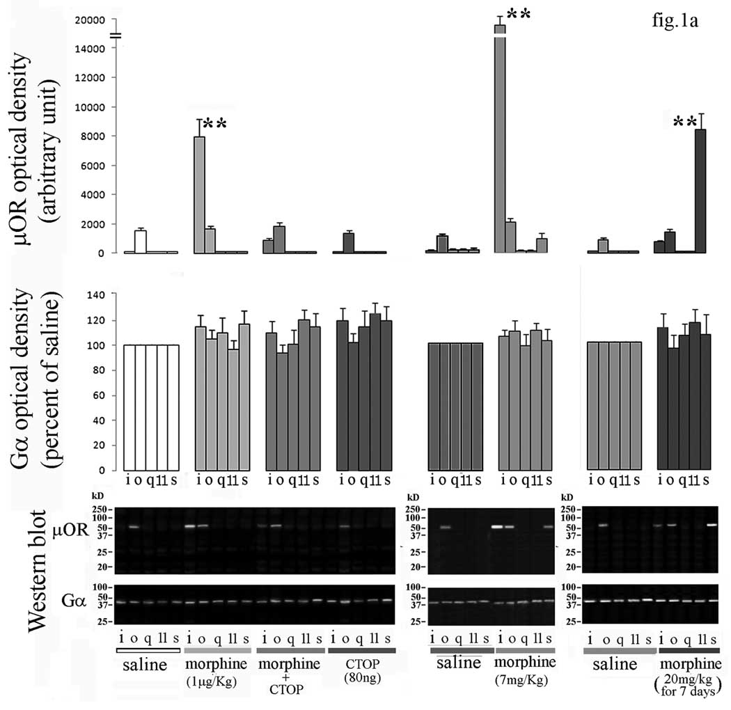
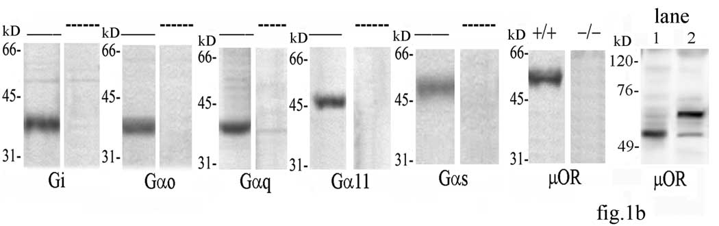
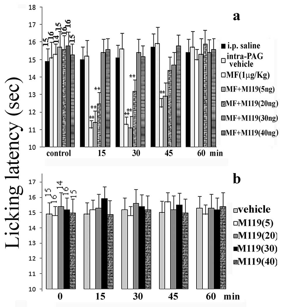
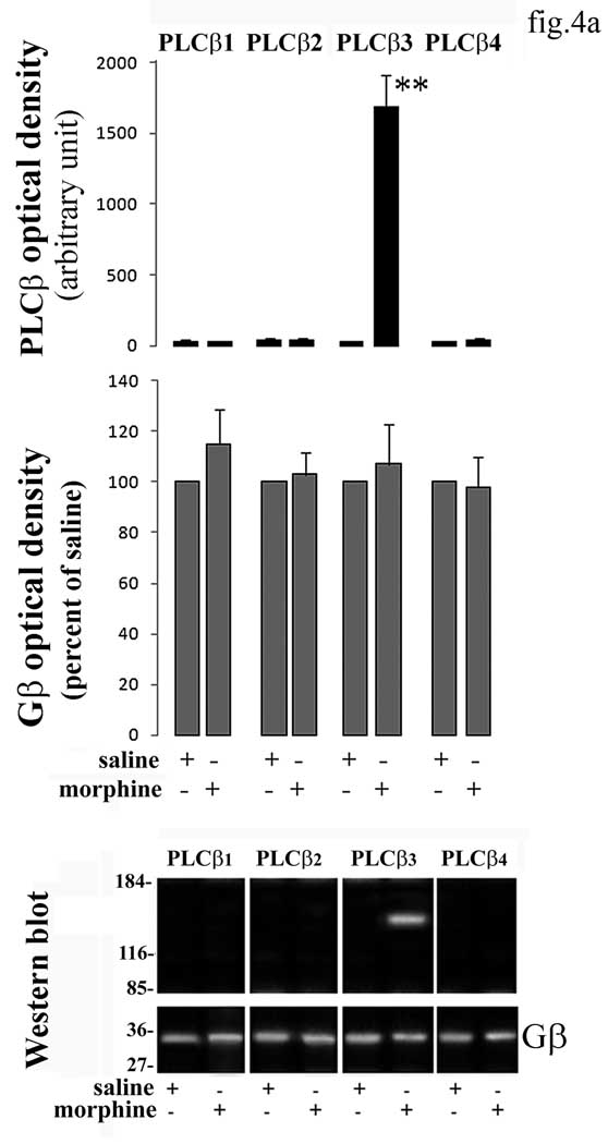
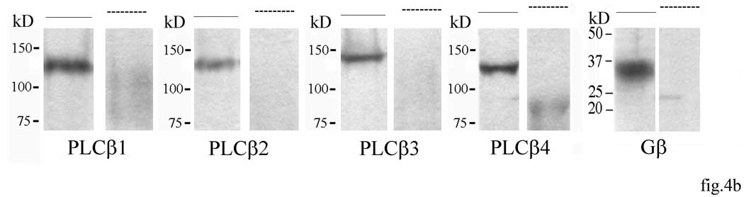
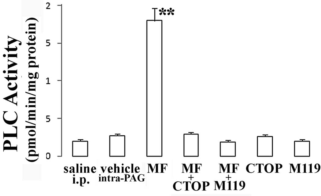
Although alterations in micro-opioid receptor (microOR) signaling mediate excitatory effects of opiates in opioid tolerance, the molecular mechanism for the excitatory effect of acute low dose morphine, as it relates to microOR coupling, is presently unknown. A pronounced coupling of microOR to the alpha subunit of G inhibitory protein emerged in periaqueductal gray (PAG) from mice systemically administered with morphine at a dose producing acute thermal hyperalgesia. This coupling was abolished in presence of the selective microOR antagonist d-Phe-Cys-Tyr-d-Trp-Orn-Thr-Pen-Thr-NH(2) administered at the PAG site, showing that the low dose morphine effect is triggered by microOR activated G inhibitory protein at supraspinal level. When Gbetagamma downstream signalling was blocked by intra-PAG co-administration of 2-(3,4,5-trihydroxy-6-oxoxanthen-9-yl)cyclohexane-1-carboxylic acid, a compound that inhibits Gbetagamma dimer-dependent signaling, a complete prevention of low dose morphine induced acute thermal hyperalgesia was obtained. Phospholipase C beta3, an enzyme necessary to morphine hyperalgesia, was revealed to be associated with Gbetagamma in PAG. Although opioid administration induces a shift in microOR-G protein coupling from Gi to Gs after chronic administration, our data support that this condition is not realized in acute treatment providing evidence that a separate molecular mechanism underlies morphine induced acute excitatory effect.


Figures

Figure 1
Figure 1
(a) G protein-μOR coupling in PAG from mice treated with morphine- A representative Western blots of the presence of μOR protein in immunoprecipitates of i, o, q, 11, and s subunits of Gα protein in PAG from mice submitted to acute or chronic morphine treatments in presence or absence of CTOP is shown at the bottom of the figure. The blots stripped and reprobed with antibodies against the above G proteins, are shown for the different treatments. Band optical density for Gα protein subunits after different treatments is represented at the middle of the figure. Each bar represents the mean density of each Gα subunit obtained from three independent experiments and expressed as percent of corresponding saline. Statistics were applied to the raw data prior to transformation to percent. Mean value of μOR density detected in immunoprecipitates of considered Gα subunits is represented at the top of the figure. Single values were normalized to surrounding background and expressed as arbitrary units. **=α<0.01 significant difference in comparison with corresponding saline value. Vertical lines represent S.E.M. (b) Specificity assay of anti-G protein and anti-μOR antibodies- Homogenates from whole mouse brain were submitted to Western blotting after incubation with the proper antibody in presence or absence of respective immunogen sequence in excess as shown in the figure. Western blotting was performed on extracts of brain tissue from normal (+/+) and μOR knockout (−/−) mice with anti-μOR antibody. Molecular weight of μOR immunoprecipitated by the anti-μOR antibody after elution with an acidic (lane 1) versus a neutral (lane 2) buffer is reported in the last column. —=not preabsorbed; ----=preabsorbed.
Figure 1
Figure 1
(a) G protein-μOR coupling in PAG from mice treated with morphine- A representative Western blots of the presence of μOR protein in immunoprecipitates of i, o, q, 11, and s subunits of Gα protein in PAG from mice submitted to acute or chronic morphine treatments in presence or absence of CTOP is shown at the bottom of the figure. The blots stripped and reprobed with antibodies against the above G proteins, are shown for the different treatments. Band optical density for Gα protein subunits after different treatments is represented at the middle of the figure. Each bar represents the mean density of each Gα subunit obtained from three independent experiments and expressed as percent of corresponding saline. Statistics were applied to the raw data prior to transformation to percent. Mean value of μOR density detected in immunoprecipitates of considered Gα subunits is represented at the top of the figure. Single values were normalized to surrounding background and expressed as arbitrary units. **=α<0.01 significant difference in comparison with corresponding saline value. Vertical lines represent S.E.M. (b) Specificity assay of anti-G protein and anti-μOR antibodies- Homogenates from whole mouse brain were submitted to Western blotting after incubation with the proper antibody in presence or absence of respective immunogen sequence in excess as shown in the figure. Western blotting was performed on extracts of brain tissue from normal (+/+) and μOR knockout (−/−) mice with anti-μOR antibody. Molecular weight of μOR immunoprecipitated by the anti-μOR antibody after elution with an acidic (lane 1) versus a neutral (lane 2) buffer is reported in the last column. —=not preabsorbed; ----=preabsorbed.
Figure 2
Figure 2
Effect of M119 coadministration on morphine induced hyperalgesic response-Licking latencies were measured before and after (15, 30, 45 and 60 min) i.p. morphine administration (1µg/Kg) in presence or absence of M119 intra-PAG coadministration at different doses (a). Licking latencies measured after M119 intra-PAG administration to mice are represented in (b). Each value represents the mean ± S.E.M. of licking latencies. Vertical bars represent S.E.M. ; **=α<0.01 significant difference in comparison with corresponding basal value. MF= morphine. The number of animals used for each experimental condition is reported at the top of control bars.
Figure 3
Figure 3
(a) Morphine and M119 administration do not induce any significant difference with respect to saline or vehicle on inspection activity and spontaneous mobility evaluated in the mouse hole board test. (b) Lack of effect of morphine and M119 administration on motor coordination evaluated in the mouse rota rod test. Vertical lines represent S.E.M.
Figure 4
Figure 4
(a) Representative blot of co-immunoprecipitation of Gβ proteins with PLCβ1–4 in PAG from low dose morphine administered mice is shown in figure. The blots stripped and reprobed with Gβ antibody are shown at the bottom of the figure. Band optical density for Gβ protein subunits after saline or morphine is represented in the middle of the figure. Each bar represents the mean density of Gβ subunit obtained from three independent experiments and expressed as percent of corresponding saline. Statistics was applied to the raw data prior to transformation to percent. Mean value of PLCβ subunit density detected in immunoprecipitates of Gβ is represented at the top of the figure. Single values were normalized to surrounding background and expressed as arbitrary units. **=α<0.01 significant difference in comparison with corresponding saline value. Vertical lines represent S.E.M. (b) Specificity assay of anti-PLCβ1–4 and anti-Gβ protein antibodies- Western blot of whole mouse brain tissue preincubated with the proper antibody in presence or absence of respective immunogen sequence in excess are shown in figure.
Figure 4
Figure 4
(a) Representative blot of co-immunoprecipitation of Gβ proteins with PLCβ1–4 in PAG from low dose morphine administered mice is shown in figure. The blots stripped and reprobed with Gβ antibody are shown at the bottom of the figure. Band optical density for Gβ protein subunits after saline or morphine is represented in the middle of the figure. Each bar represents the mean density of Gβ subunit obtained from three independent experiments and expressed as percent of corresponding saline. Statistics was applied to the raw data prior to transformation to percent. Mean value of PLCβ subunit density detected in immunoprecipitates of Gβ is represented at the top of the figure. Single values were normalized to surrounding background and expressed as arbitrary units. **=α<0.01 significant difference in comparison with corresponding saline value. Vertical lines represent S.E.M. (b) Specificity assay of anti-PLCβ1–4 and anti-Gβ protein antibodies- Western blot of whole mouse brain tissue preincubated with the proper antibody in presence or absence of respective immunogen sequence in excess are shown in figure.
Figure 5
Figure 5
PLC activity present in anti-Gβ immunoprecipitates from PAG of mice previously administered with morphine (1 µg/Kg) in presence or absence of CTOP (80ng) or M119(40ng). Each bar represents PLC activity obtained from three independent experiments under different treatment conditions. Immune complex PLC activity was measured and expressed as pmol Ins(1,4,5)P3 product formed/min/mg protein. Vertical lines represent S.E.M.;**=α<0.01.

Similar articles

Cited by

References

    1. Abbott FV, Franklin KB, Connell B. The stress of a novel environment reduces formalin pain: possible role of serotonin. Eur J Pharmacol. 1986;126:141–144. - PubMed
    1. Allan AM, Weeber EJ, Savage DD, Caldwell KK. Effects of prenatal ethanol exposure on phospholipase C-beta 1 and phospholipase A2 in hippocampus and medial frontal cortex of adult rat offspring. Alcohol Clin Exp Res. 1997;21:1534–1541. - PubMed
    1. Askari N, Mahboudi F, Haeri-Rohani A, Kazemi B, Sarrami R, Edalat R, Ahmadiani A. Effects of single administration of morphine on G-protein mRNA level in the presence and absence of inflammation in the rat spinal cord. Scand J Immunol. 2008;67:47–52. - PubMed
    1. Bianchi E, Lehmann D, Vivoli E, Norcini M, Ghelardini C. Involvement of PLC-{beta}3 in the effect of morphine on memory retrieval in passive avoidance task. J Psychopharmacol. 2009 Mar 12; [Epub ahead of print] - PubMed
    1. Bonacci TM, Mathews JL, Yuan C, Lehmann DM, Malik S, Wu D, Font JL, Bidlack JM, Smrcka AV. Differential targeting of Gbetagamma-subunit signaling with small molecules. Science. 2006;312:443–446. - PubMed

Publication types

MeSH terms