. 2009 Aug 5:9:29.
doi: 10.1186/1472-6882-9-29.

Inhibition of HIV-1 entry by extracts derived from traditional Chinese medicinal herbal plants

Affiliations

Inhibition of HIV-1 entry by extracts derived from traditional Chinese medicinal herbal plants

In-Woo Park et al. BMC Complement Altern Med. .

Abstract

Background: Highly active anti-retroviral therapy (HAART) is the current HIV/AIDS treatment modality. Despite the fact that HAART is very effective in suppressing HIV-1 replication and reducing the mortality of HIV/AIDS patients, it has become increasingly clear that HAART does not offer an ultimate cure to HIV/AIDS. The high cost of the HAART regimen has impeded its delivery to over 90% of the HIV/AIDS population in the world. This reality has urgently called for the need to develop inexpensive alternative anti-HIV/AIDS therapy. This need has further manifested by recent clinical trial failures in anti-HIV-1 vaccines and microbicides. In the current study, we characterized a panel of extracts of traditional Chinese medicinal herbal plants for their activities against HIV-1 replication.

Methods: Crude and fractionated extracts were prepared from various parts of nine traditional Chinese medicinal herbal plants in Hainan Island, China. These extracts were first screened for their anti-HIV activity and cytotoxicity in human CD4+ Jurkat cells. Then, a single-round pseudotyped HIV-luciferase reporter virus system (HIV-Luc) was used to identify potential anti-HIV mechanisms of these extracts.

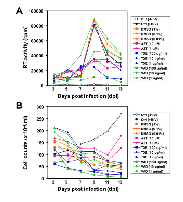
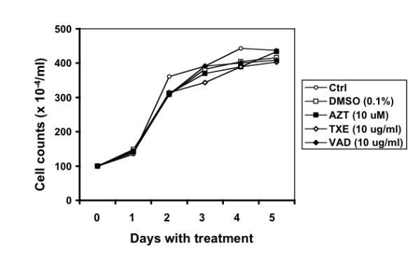
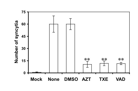
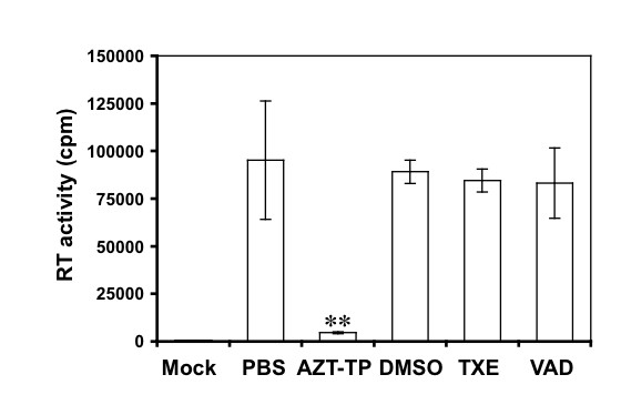
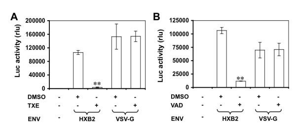
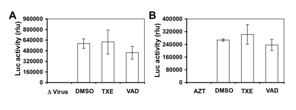
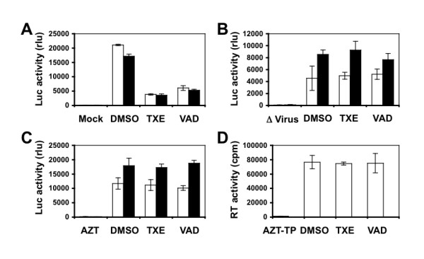
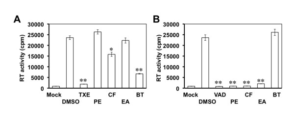
Results: Two extracts, one from Euphorbiaceae, Trigonostema xyphophylloides (TXE) and one from Dipterocarpaceae, Vatica astrotricha (VAD) inhibited HIV-1 replication and syncytia formation in CD4+ Jurkat cells, and had little adverse effects on host cell proliferation and survival. TXE and VAD did not show any direct inhibitory effects on the HIV-1 RT enzymatic activity. Treatment of these two extracts during the infection significantly blocked infection of the reporter virus. However, pre-treatment of the reporter virus with the extracts and treatment of the extracts post-infection had little effects on the infectivity or gene expression of the reporter virus.

Conclusion: These results demonstrate that TXE and VAD inhibit HIV-1 replication likely by blocking HIV-1 interaction with target cells, i.e., the interaction between gp120 and CD4/CCR5 or gp120 and CD4/CXCR4 and point to the potential of developing these two extracts to be HIV-1 entry inhibitors.


Figures

Figure 1
Figure 1
Effects of the extracts from TXE and VAD on HIV replication and cell survival. Jurkat cells were infected with HIV-1 HXB2 and then exposed to the extracts 24 hr post infection. Fresh extracts were added every other day. Meanwhile, culture supernatants were collected for the RT activity assay (A), and aliquots of cells were stained with trypan blue dye and counted for viable cells (B). DMSO was the solvent of the extracts and included as a negative control, while AZT was included as a positive control. In addition, Jurkat cells without HIV infection [Ctrl, (-HIV)] and Jurkat cells with HIV infection but without any treatments [Ctrl, (+HIV)] were also included. These data were representative of three independent experiments.
Figure 2
Figure 2
Effects of the extracts on cell proliferation and survival. Jurkat cells were exposed to the extracts for various lengths of time as indicated. Fresh extracts were added every other day. Cells without any treatments, treated with DMSO, or AZT were included as controls. Viable cells were determined using the trypan blue dye staining. These data were representative of three independent experiments.
Figure 3
Figure 3
Effect of the extracts on syncytia formation in HIV-1-infected Jurkat cells. Jurkat cells were infected with HIV-1 and then exposed to the extracts at 10 μg/ml, 0.1% DMSO, or 5 μM AZT. Syncytia in each of these treatments were counted from 4 random fields from each one of the triplicate samples under a light microscope over the course of 2 weeks infection. The data represented the number of syncytia at day 7 post infection when the maximal number of syncytia was recorded in the infections receiving no treatments (None) or DMSO. Jurkat cells without HIV-1 infection were included as a control (Mock). The data were mean ± SEM of triplicate experiments.
Figure 4
Figure 4
Direct effects of the extracts on the RT activity. HIV-1 virions were assayed for their RT activity in the presence of the extracts at 10 μg/ml. AZT-TP (5 μM) was included as a positive control. DMSO (0.1%) and phosphate-buffered saline (PBS) were included as the solvent control for the extracts and AZT, respectively. The RT reaction without any input HIV-1 virions was also included as an assay control. The data were mean ± SEM of triplicate experiments.
Figure 5
Figure 5
Effects of the extracts on HIV-1 entry. U87.CD4.CXCR4 cells were treated with TXE (A) or VAD (B) at a concentration of 10 μg/ml for 30 min and then infected with HIV-Luc viruses pseudotyped T-tropic HIV-1 HXB2 envelope (HXB2) or without envelope (-) for 2 hr. Forty-eight hours post infection, cells were harvested for the Luc activity assay. HIV-Luc viruses pseudotyped with VSV-G envelope (VSV-G) were included as a control. DMSO (0.1%) was also included as a solvent control for the extracts. The data were mean ± SEM of triplicate experiments.
Figure 6
Figure 6
Effects of the extracts on HIV-1 and HIV-1 gene expression. A. HIV-Luc viruses pseudotyped T-tropic HIV-1 HXB2 envelope (HXB2) were incubated with 10 μg/ml extracts for 2 hr and then used to infect U87.CD4.CXCR4 cells. Cells were harvested 48 hr for the Luc activity assay 48 hr after infection. Infection with heat-inactivated HIV-Luc/HXB2 viruses (Δ Virus) was included as the control. B. U87.CD4.CXCR4 cells were infected with HIV-Luc viruses pseudotyped T-tropic HIV-1 HXB2 envelope (HXB2) or without envelope (-) for 2 hr and then removed of the remaining input viruses by repeated washes with fresh medium. Then, the infected cells were cultured for 48 hr in the presence of the extracts (10 μg/ml) and then harvested for the Luc activity assay. DMSO (0.1%) was also included as a solvent control for the extracts, while 0.5 μM AZT was included as a positive control. The data were mean ± SEM of triplicate experiments.
Figure 7
Figure 7
Effects of the extracts on primary HIV-1 isolate 89.6. A. U87.CD4.CXCR4 and U87.CD4.CCR5 cells were first treated with 10 μg/ml extracts and then infected with HIV-Luc viruses pseudotyped with 89.6 envelope. Infection of HIV-Luc viruses without an envelope was included as the mock infection control. B. HIV-Luc viruses pseudotyped 89.6 envelope were first incubated with 10 μg/ml extracts and then used to infect U87.CD4.CXCR4 and U87.CD4.CCR5 cells. Infection with heat-inactivated HIV-Luc/89.6 viruses (Δ Virus) was included as the control. C. U87.CD4.CXCR4 and U87.CD4.CCR5 were first infected with HIV-Luc viruses pseudotyped with 89.6 envelope and then treated with 10 μg/ml extracts or 5 μM AZT. A-C: open bar for U87.CD4.CXCR4 cells; closed bar for U87.CD4.CCR5 cells. D. HIV-Luc viruses pseudotyped with 89.6 envelope were directly treated with 10 μg/ml extracts or 5 μM AZT-TP, the RT activity was determined. The data were mean ± SEM of triplicate experiments.
Figure 8
Figure 8
Anti-HIV components of TXE and VAD extracts. Jurkat cells were infected with HIV-1 HXB2 and then exposed to 10 μg/ml TXE (A), VAD (B), or each of its partition subfractions from petroleum ether (PE), chloroform (CF), ethyl acetate (EA) and n-butanol (BT) 24 hr post infection. Fresh extracts or subfraction were added every other day. Meanwhile, culture supernatants were collected for the RT activity assay, and aliquots of cells were stained with trypan blue dye and counted for viable cells. DMSO was the solvent of the extracts and subfractions and included as a vehicle control. The RT data from the supernatants collected at day 9 of the peak viral replication were presented. Extracts and their subfractions showed no apparent cytotoxic effects on the cells. The data were mean ± SEM of triplicate experiments.

Similar articles

Cited by

References

    1. Barre-Sinoussi F, Chermann JC, Rey F, Nugeyre MT, Chamaret S, Gruest J, Dauguet C, Axler-Blin C, Vezinet-Brun F, Rouzioux C, et al. Isolation of a T-lymphotropic retrovirus from a patient at risk for acquired immune deficiency syndrome (AIDS) Science. 1983;220:868–871. doi: 10.1126/science.6189183. - DOI - PubMed
    1. Broder S, Gallo RC. A pathogenic retrovirus (HTLV-III) linked to AIDS. N Engl J Med. 1984;311:1292–1297. - PubMed
    1. Deen KC, McDougal JS, Inacker R, Folena-Wasserman G, Arthos J, Rosenberg J, Maddon PJ, Axel R, Sweet RW. A soluble form of CD4 (T4) protein inhibits AIDS virus infection. Nature. 1988;331:82–84. doi: 10.1038/331082a0. - DOI - PubMed
    1. Eckert DM, Kim PS. Mechanisms of viral membrane fusion and its inhibition. Annu Rev Biochem. 2001;70:777–810. doi: 10.1146/annurev.biochem.70.1.777. - DOI - PubMed
    1. Platt EJ, Durnin JP, Kabat D. Kinetic factors control efficiencies of cell entry, efficacies of entry inhibitors, and mechanisms of adaptation of human immunodeficiency virus. J Virol. 2005;79:4347–4356. doi: 10.1128/JVI.79.7.4347-4356.2005. - DOI - PMC - PubMed

Publication types