. 2009 Aug;10(2):119-30.
doi: 10.1016/j.cmet.2009.06.012.

Discovery of genes essential for heme biosynthesis through large-scale gene expression analysis

Affiliations

Discovery of genes essential for heme biosynthesis through large-scale gene expression analysis

Roland Nilsson et al. Cell Metab. 2009 Aug.

Abstract

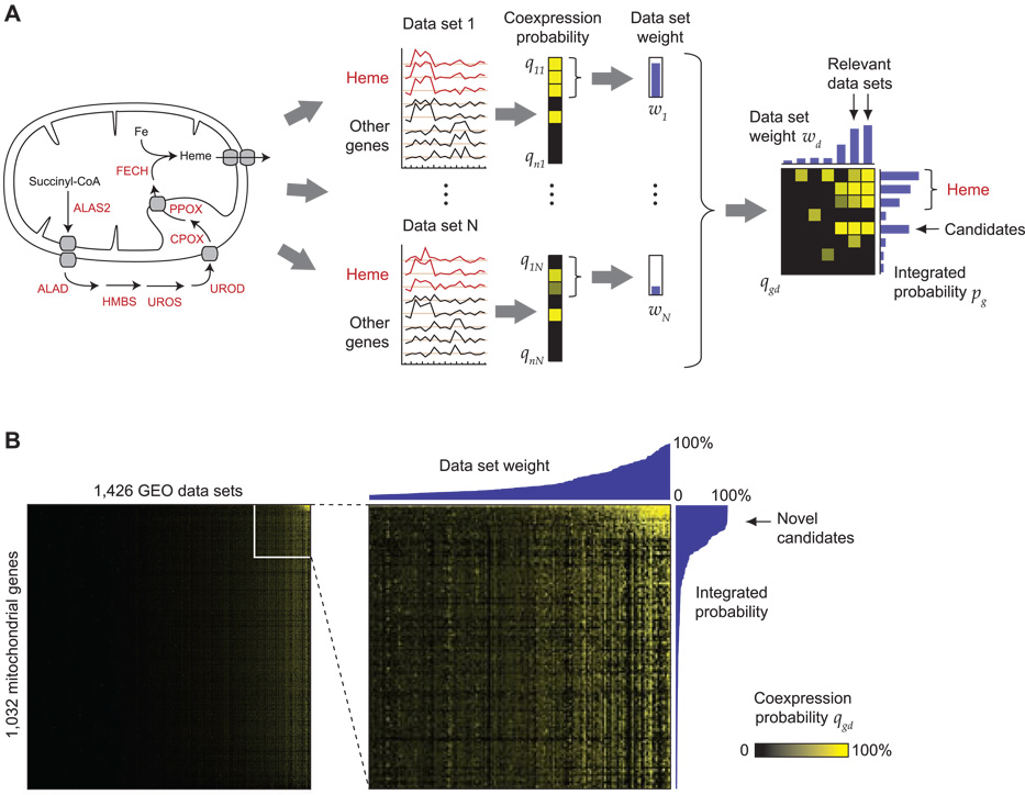
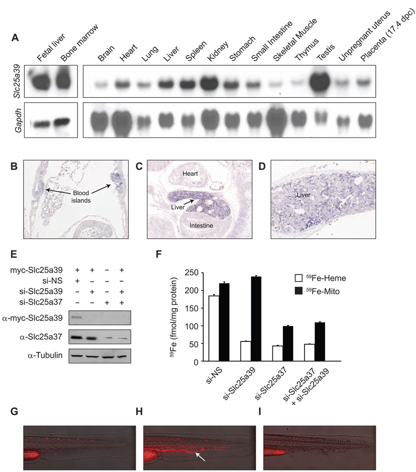
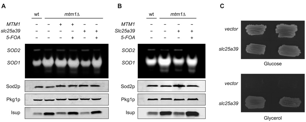
Heme biosynthesis consists of a series of eight enzymatic reactions that originate in mitochondria and continue in the cytosol before returning to mitochondria. Although these core enzymes are well studied, additional mitochondrial transporters and regulatory factors are predicted to be required. To discover such unknown components, we utilized a large-scale computational screen to identify mitochondrial proteins whose transcripts consistently coexpress with the core machinery of heme biosynthesis. We identified SLC25A39, SLC22A4, and TMEM14C, which are putative mitochondrial transporters, as well as C1orf69 and ISCA1, which are iron-sulfur cluster proteins. Targeted knockdowns of all five genes in zebrafish resulted in profound anemia without impacting erythroid lineage specification. Moreover, silencing of Slc25a39 in murine erythroleukemia cells impaired iron incorporation into protoporphyrin IX, and vertebrate Slc25a39 complemented an iron homeostasis defect in the orthologous yeast mtm1Delta deletion mutant. Our results advance the molecular understanding of heme biosynthesis and offer promising candidate genes for inherited anemias.


Figures

Figure 1
Figure 1
Identifying novel candidate genes for heme biosynthesis using expression screening. (A) The eight known enzymes of heme biosynthesis pathway (left) defines the query pathway. Using a rank-based statistic, each gene g is assigned a probability of coexpression qgd in each microarray data set d (black/yellow columns). Data sets where the heme biosynthesis enzymes are themselves tightly coexpressed are assigned larger weights wd (blue vertical bars), which are then used to integrate the coexpression information from all data sets (black/yellow matrix, right) into a final probability pg (blue horizontal bars). (B) The coexpression matrix for the 1,426 data sets used in this study (columns), over 1,032 mitochondrial genes (rows). Yellow color indicates strong coexpression. Right, a magnified portion of this matrix, with data set weights (top) and integrated probabilities (right).
Figure 2
Figure 2
Microarray datasets where the heme biosynthetic pathway is regulated. (A) Time-series gene expression of heme biosynthesis enzymes and five selected candidates (see text) during erythroid differentiation. (B) Gene expression in Nix−/− and wild-type (WT) mouse spleen. (C) Gene expression in Rb−/− and wild-type (WT) mouse fetal liver. (D) Gene expression in a panel of mouse tissues. Red color indicates high expression; blue, low expression; gray color, missing values. GSE, NCBI Gene Expression Omnibus accession number.
Figure 3
Figure 3
Expression of candidate genes in zebrafish blood islands. Whole embryo in situ hybridization was performed on embryos at 24hpf. (A) Slc4a1 was used as control to delineate the intermediate cell mass (ICM, indicated by arrows). (B) Cloche (clo) embryos were used to show specificity of the hybridizations. (C–G) Candidates identified in this study.
Figure 4
Figure 4
Morpholino knockdown of candidate genes in zebrafish results in profound anemia. WT zebrafish embryos were injected at the 1-cell stage with the respective morpholinos and stained at 48hpf with o-dianisidine to detect hemoglobinized cells. (A) Uninjected (wt) embryos show normal hemoglobinization as indicated by dark-brown staining on the yolk sac (arrow). (B–G) Morpholino-injected embryos. Accurate morpholino gene targeting was verified by RT-PCR (slc25a39, slc22a4, slc22a5, tmem14c, and c1orf69) or real-time quantitative RT-PCR (isca1) on cDNA from uninjected (wt) or morpholino-injected (mo) embryos. β-actin (actb) was used as a control for off-target effects in the RT-PCR. For RT-PCR, (ctrl) indicates no cDNA template control. For real-time quantitative RT-PCR, (ctrl mo) indicates embryos injected with a standard control morpholino.
Figure 5
Figure 5
Mouse Slc25a39 is expressed in hematopoietic tissues and is important for heme biosynthesis. (A) Mouse tissue northern blot analysis of Slc25a39 expression. (B) Mouse Slc25a39 transcripts are localized to blood islands of the yolk sac at early somite stages (E8.5, arrows). (C) and (D) Slc25a39 transcripts accumulate most abundantly in the liver where expression is heterogeneous (E12.5). (E) and (F) MEL cells were differentiated for two days in media containing 1.5% DMSO prior to (E) transient transfection with myc-tagged Slc25a39 or (F) silencing of Slc25a39 (si-Slc25a39) and/or Slc25a37 (si-Slc25a37) using siRNA oligos. Assays were performed after two additional days of differentiation. si-NS indicates silencing using non-specific control oligos. (E) Representative western blot using anti-myc (α-myc-Slc25a39) and anti-Slc25a37 antibodies. Equal loading was verified by anti-tubulin. (F) MEL cells were metabolically labeled with 59Fe conjugated to transferrin, and total mitochondrial iron (59Fe-Mito) and iron in heme (59Fe-Heme) were determined. Results shown are from two independent experiments assayed in duplicate; error bars denote standard deviation. (G) (H) and (I) Representative photos of (G) an uninjected, wild-type control zebrafish embryo, (H) a zebrafish embryo injected with a ferrochelatase-specific morpholino, or (H) with a slc25a39-specific morpholino (I). Arrow indicates porphyric red blood cells in circulation.
Figure 6
Figure 6
Vertebrate SLC25A39 is the ortholog of yeast MTM1 and is involved in mitochondrial iron homeostasis. (A) and (B) mtm1Δ strains were transformed with URA3 based plasmids for expressing (A and B) S. cerevisiae MTM1, (A) zebrafish slc25a39, or (B) mouse Slc25a39. 5-FOA indicates transformants induced to shed the plasmids by growth on 5-fluoroorotic acid. Whole cell lysates of the transformants, 5-FOA derivatives and the parental WT and mtm1Δ cells were subjected to (top) native gel electrophoresis and NBT staining for SOD (SOD2 and SOD1) activity, and (bottom) to SDS-PAGE and immunoblotting for Sod2p, Pgk1 (loading control) and Isup (sum of Isu1p and Isu2p). (C) WT strain transformed with either empty pRS426-ADH vector or zebrafish slc25A39 was subjected to MTM1 deletion and tested for mitochondrial DNA function by growth on fermentable (glucose) versus non-fermentable (glycerol) carbon sources. Results shown represent two independent colonies.

Comment in

References

    1. Ajioka RS, Phillips JD, Kushner JP. Biosynthesis of heme in mammals. Biochim Biophys Acta. 2006;1763:723–736. - PubMed
    1. Allikmets R, Raskind WH, Hutchinson A, Schueck ND, Dean M, Koeller DM. Mutation of a putative mitochondrial iron transporter gene (ABC7) in X-linked sideroblastic anemia and ataxia (XLSA/A) Hum Mol Genet. 1999;8:743–749. - PubMed
    1. Barrett T, Troup DB, Wilhite SE, Ledoux P, Rudnev D, Evangelista C, Kim IF, Soboleva A, Tomashevsky M, Edgar R. NCBI GEO: mining tens of millions of expression profiles--database and tools update. Nucleic Acids Res. 2007;35:D760–D765. - PMC - PubMed
    1. Bennett CM, Kanki JP, Rhodes J, Liu TX, Paw BH, Kieran MW, Langenau DM, Delahaye-Brown A, Zon LI, Fleming MD, et al. Myelopoiesis in the zebrafish, Danio rerio. Blood. 2001;98:643–651. - PubMed
    1. Boeke JD, Trueheart J, Natsoulis G, Fink GR. 5-Fluoroorotic acid as a selective agent in yeast molecular genetics. Methods Enzymol. 1987;154:164–175. - PubMed

Publication types

MeSH terms