

Transforming growth factor-beta1 modulates insulin-like growth factor binding protein-4 expression and proteolysis in cultured periosteal explants
- PMID: 19656700
- PMCID: PMC2844918
- DOI: 10.1016/j.ghir.2009.06.002
Transforming growth factor-beta1 modulates insulin-like growth factor binding protein-4 expression and proteolysis in cultured periosteal explants
Abstract
Objective: Periosteum is involved in bone growth and fracture healing and has been used as a cell source and tissue graft for tissue engineering and orthopedic reconstruction including joint resurfacing. Periosteum can be induced by transforming growth factor beta (TGF-beta) or insulin-like growth factor-I (IGF-I) alone or in combination to form cartilage. However, little is known about the interaction between IGF and TGF-beta signaling during periosteal chondrogenesis. The purpose of this study was to determine the effect of TGF-beta1 on IGF binding protein-4 (IGFBP-4) and the IGFBP-4 protease pregnancy-associated plasma protein-A (PAPP-A) expression in cultured periosteal explants.
Design: Periosteal explants from rabbits were cultured with or without TGF-beta1. IGFBP-4 and PAPP-A mRNA levels were determined by real-time quantitative PCR. Conditioned medium was analyzed for IGFBP-4 and PAPP-A protein levels and IGFBP-4 protease activity.
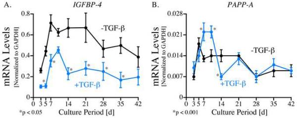
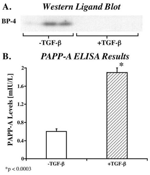
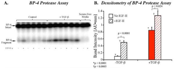
Results: TGF-beta1-treated explants contained lower IGFBP-4 mRNA levels throughout the culture period with a maximum reduction of 70% on day 5 of culture. Lower levels of IGFBP-4 protein were also detected in the conditioned medium from TGF-beta1-treated explants. PAPP-A mRNA levels were increased 1.6-fold, PAPP-A protein levels were increased threefold, and IGFBP-4 protease activity was increased 8.5-fold between 7 and 10days of culture (the onset of cartilage formation in this model) in conditioned medium from TGF-beta1-treated explants.
Conclusions: This study demonstrates that TGF-beta1 modulates the expression of IGFBP-4 and PAPP-A in cultured periosteal explants.
Copyright (c) 2009 Elsevier Ltd. All rights reserved.
Figures
References
-
- O'Driscoll SW, Salter RB. The induction of neochondrogenesis in free intra-articular periosteal autografts under the influence of continuous passive motion. An experimental investigation in the rabbit. J Bone Joint Surg. 1984;66A:1248–1257. - PubMed
-
- O'Driscoll SW, Keeley FW, Salter RB. Durability of regenerated articular cartilage produced by free autogenous periosteal grafts in major full-thickness defects in joint surfaces under the influence of continuous passive motion. A follow-up report at one year. J Bone Joint Surg. 1988;70A:595–606. - PubMed
-
- O'Driscoll SW, Salter RB. The repair of major osteochondral defects in joint surfaces by neochondrogenesis using autogenous osteoperiosteal grafts stimulated by continuous passive motion. An experimental investigation in the rabbit. Clin Orthop. 1986;208:131–140. - PubMed
-
- O'Driscoll SW, Keeley FW, Salter RB. The chondrogenic potential of free autogenous periosteal grafts for biological resurfacing of major full-thickness defects in joint surfaces under the influence of continuous passive motion. An experimental investigation in the rabbit. J Bone Joint Surg. 1986;68A:1017–1035. - PubMed
-
- O'Driscoll SW. Articular cartilage regeneration using periosteum. Clin Orthop. 1999;367(Suppl):186–203. - PubMed
Publication types
MeSH terms
Substances
Grants and funding
LinkOut - more resources
Full Text Sources
Miscellaneous
