Review
. 2009 Oct 6;6 Suppl 5(Suppl 5):S575-85.
doi: 10.1098/rsif.2009.0167.focus. Epub 2009 Aug 5.

Large facilities and the evolving ribosome, the cellular machine for genetic-code translation

Affiliations
Review

Large facilities and the evolving ribosome, the cellular machine for genetic-code translation

Ada Yonath. J R Soc Interface. .

Abstract

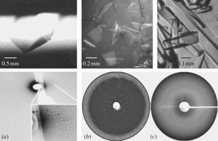
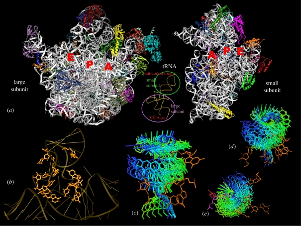
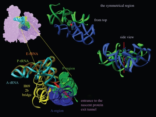
Well-focused X-ray beams, generated by advanced synchrotron radiation facilities, yielded high-resolution diffraction data from crystals of ribosomes, the cellular nano-machines that translate the genetic code into proteins. These structures revealed the decoding mechanism, localized the mRNA path and the positions of the tRNA molecules in the ribosome and illuminated the interactions of the ribosome with initiation, release and recycling factors. They also showed that the ribosome is a ribozyme whose active site is situated within a universal symmetrical region that is embedded in the otherwise asymmetric ribosome structure. As this highly conserved region provides the machinery required for peptide bond formation and for ribosome polymerase activity, it may be the remnant of the proto-ribosome, a dimeric pre-biotic machine that formed peptide bonds and non-coded polypeptide chains. Synchrotron radiation also enabled the determination of structures of complexes of ribosomes with antibiotics targeting them, which revealed the principles allowing for their clinical use, revealed resistance mechanisms and showed the bases for discriminating pathogens from hosts, hence providing valuable structural information for antibiotics improvement.


Figures

Figure 1.
Figure 1.
From poor to useful diffraction of ribosomal crystals: (a) the tip of an approximately 2 mm long crystal of B50S and its diffraction pattern obtained in 1984 at the EMBL beam line at DESY/Hamburg at 4°C. (b) Crystals of H50S and their diffraction pattern obtained in 1998 at ID13 ESRF at −180°C. Note that the diffraction extends to 2.8 Å, although the crystals are extremely thin. (c) Diffraction pattern obtained from a multi-tungsten-cluster treated T30S crystal in 1998 at 19ID/APS/ANL at −180°C.
Figure 2.
Figure 2.
The PTC and the passage of the tRNA 3′end by rotatory motion. (a) The interface faces as seen in the 3 Å structures of the two ribosomal subunits of the eubacterium D. radiodurans (rRNA is shown in silver, and each of the r-proteins is shown in a different colour). Note that these interfaces are rich in RNA. Inset: the backbone of a tRNA molecule. The circles designate the regions interacting with each of the ribosomal subunits. (b) A view into the PTC backbone. The nucleotides located in proximity to the substrates are shown in detail. (ce) Snapshots of the tRNA 3′end passage from A- to P-site, represented by the transition from the A-site aminoacylated tRNA (in blue) to the P-site (in green), obtained computationally by successive rotations by 15° each around the bond connecting the 3′end to the rest of the tRNA, with the ribosomal nucleotides that interact with the motion (in gold). (a) An orthogonal view and (d,e) two side views are shown. (e) The two flexible nucleotides (A2602 and U2585) that seem to anchor and propel this motion are shown in red and magenta, respectively.
Figure 3.
Figure 3.
The ribosomal symmetrical region and suggested proto-ribosome. In all, the region hosting A-site tRNA is shown in blue and that hosting the P-site tRNA in green. Similarly, the A-site tRNA mimic (Bashan et al. 2003) is shown in blue, and the derived P-site tRNA (by the rotatory motion) is shown in green. The imaginary symmetrical axis is shown in red. Top left: the symmetrical region within the ribosome and its details. The A-region is shown in blue, the P-region in green and the non-symmetrical extensions are shown as pink dots on green ‘ropes’. A- and P-site tRNAs are shown in cyan and green-yellow. A zoom into the symmetrical region is shown in bottom left. The bridge to the decoding centre and A-, P- and E-site tRNA molecules were docked, based on Yusupov et al. (2001).

References

    1. Agmon I., et al. 2003. On peptide bond formation, translocation, nascent protein progression and the regulatory properties of ribosomes. Eur. J. Biochem. 270, 2543–2556. (10.1046/j.1432-1033.2003.03634.x) - DOI - PubMed
    1. Agmon I., Bashan A., Zarivach R., Yonath A. 2005. Symmetry at the active site of the ribosome: structural and functional implications. Biol. Chem. 386, 833–844. (10.1515/BC.2005.098) - DOI - PubMed
    1. Agmon I., Bashan A., Yonath A. 2006. On ribosome conservation and evolution. Isr. J. Ecol. Evol. 52, 359–379. (10.1560/IJEE_52_3-4_359) - DOI
    1. Agmon I., Davidovich C., Bashan A., Yonath A. 2009. Identification of the prebiotic translation apparatus within the contemporary ribosome. See http://precedings.nature.com/documents/2921/version/1.
    1. Amit M., Berisio R., Baram D., Harms J., Bashan A., Yonath A. 2005. A crevice adjoining the ribosome tunnel: hints for cotranslational folding. FEBS Lett. 579, 3207–3213. (10.1016/j.febslet.2005.03.023) - DOI - PubMed

Publication types

MeSH terms