. 2009 Oct;83(20):10761-9.
doi: 10.1128/JVI.00770-09. Epub 2009 Aug 5.

Activation of MDA5 requires higher-order RNA structures generated during virus infection

Affiliations

Activation of MDA5 requires higher-order RNA structures generated during virus infection

Andreas Pichlmair et al. J Virol. 2009 Oct.

Abstract

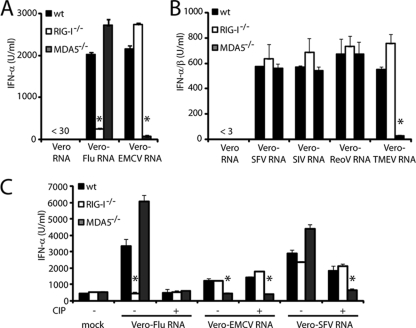
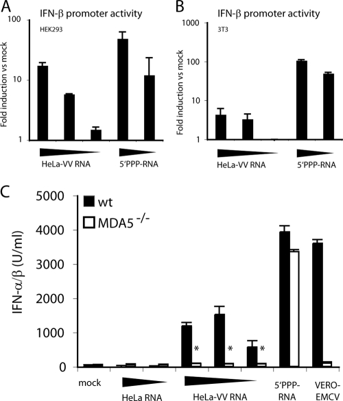
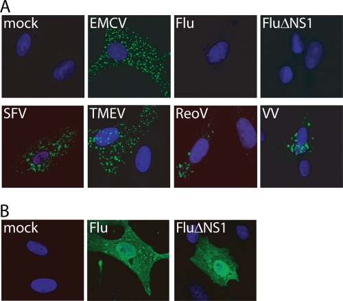
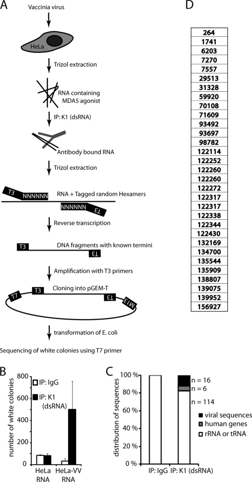
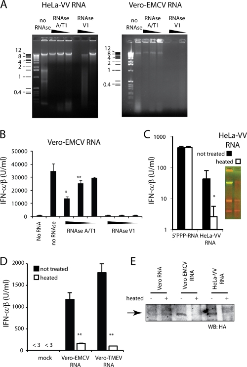
Recognition of virus presence via RIG-I (retinoic acid inducible gene I) and/or MDA5 (melanoma differentiation-associated protein 5) initiates a signaling cascade that culminates in transcription of innate response genes such as those encoding the alpha/beta interferon (IFN-alpha/beta) cytokines. It is generally assumed that MDA5 is activated by long molecules of double-stranded RNA (dsRNA) produced by annealing of complementary RNAs generated during viral infection. Here, we used an antibody to dsRNA to show that the presence of immunoreactivity in virus-infected cells does indeed correlate with the ability of RNA extracted from these cells to activate MDA5. Furthermore, RNA from cells infected with encephalomyocarditis virus or with vaccinia virus and precipitated with the anti-dsRNA antibody can bind to MDA5 and induce MDA5-dependent IFN-alpha/beta production upon transfection into indicator cells. However, a prominent band of dsRNA apparent in cells infected with either virus does not stimulate IFN-alpha/beta production. Instead, stimulatory activity resides in higher-order structured RNA that contains single-stranded RNA and dsRNA. These results suggest that MDA5 activation requires an RNA web rather than simply long molecules of dsRNA.


Figures

FIG. 1.
FIG. 1.
RNAs from virus-infected cells act as RLR agonists. (A and B) MEFs of the indicated genotype were transfected with 0.2 μg of the indicated RNA, and accumulation of IFN-α in supernatants was measured by ELISA after overnight incubation. (B) MEFs of the indicated genotype were transfected with 0.2 μg of RNA from virus-infected cells. After overnight incubation, IFN-α/β in the supernatant was measured on indicator cells containing an ISRE-Luc reporter. (C) RNA from infected cells was treated with CIP (+) or buffer only (−) before transfection. IFN-α was measured by ELISA. Graphs show one of at least three representative experiments. Error bars show standard deviation of triplicate (A and C) or quadruplicate (B) measurements. Asterisks indicate a P value of <0.01 compared to wild-type (wt) control MEFs.
FIG. 2.
FIG. 2.
Transfection of RNA from virus-infected cells generates progeny virus. (A and B) 3T3 cells were left untreated or were pretreated with ribavirin for 1 h and transfected with Vero-EMCV RNA (0.2 μg in A; 1 and 0.2 μg in B) or infected with EMCV (MOI of 1). (A) Accumulation of infectious virus particles quantified by 50% tissue culture infective dose. (B) IFN-α was measured after overnight incubation. ELISA shows average of triplicate measurements + standard deviation. (C and D) Quantitative reverse transcription-PCR analysis for EMCV RNA (C) and IFN-β mRNA (D) in cells treated with CHX for 30 min and subsequently infected with EMCV or transfected with 0.2 μg of Vero-EMCV RNA for 4 h. Bars show averages of duplicate measurements + standard deviations. Graphs show one of two (A and B) or three representative experiments (C and D). Error bars show standard deviations of quadruplicate (A), triplicate (B), or duplicate (C and D) measurements. Asterisks indicate a P value of <0.001 compared to dimethyl sulfoxide (DMSO) treatment.
FIG. 3.
FIG. 3.
RNA from VV-infected cells stimulates MDA5. HEK293 (A) or 3T3 (B) cells were transfected with reporter constructs for IFN-β-luciferase and control plasmid pRL-TK and subsequently stimulated with HeLa-VV RNA (1, 0.2, and 0.04 μg) or control 5′ PPP-RNA (0.2 and 0.04 μg), respectively. Graphs show relative activation of the IFN-β promoter. Data are the average of duplicate measurements ± standard deviation. (C) MEFs of the indicated genotype were transfected with HeLa-RNA (1 and 0.2 μg), HeLa-VV RNA (1, 0.2, and 0.04 μg), Vero-EMCV RNA (0.2 μg), or with control 5′ PPP-RNA (0.2 μg). The graph shows the accumulation of IFN-α/β in supernatants after overnight incubation. *, P < 0.001 compared to wild-type (wt) MEFs. Data are average ± standard deviations of duplicate (A and B) or quadruplicate (C) measurements from one experiment repeated three times.
FIG. 4.
FIG. 4.
MDA5-activating viruses generate immunodetectable dsRNA. (A) Vero cells were infected with the indicated virus (MOI of 0.5 to 1) for 8 h (EMCV, TMEV, and SFV) or 16 h (influenza virus (Flu), FluΔNS1, and ReoV). HeLa cells were infected with VV (MOI of 0.5) for 16 h. Cells were fixed and stained with the K1 (dsRNA) MAb (green) and DAPI (blue). (B) Influenza virus-infected cells stained with anti-influenza (Flu) antibodies (green) and DAPI (blue).
FIG. 5.
FIG. 5.
K1 and MDA5 bind to the same type of RNA. (A) 293T cells were transfected with HA-tagged MDA5 and 40 h later infected with EMCV (if EMCV) or left uninfected. Eight hours later, cell lysates were prepared and used for coimmunoprecipitation experiments. Uninfected cells (lanes 5, 6, and 8) or infected cells (lane 7) were used for immunoprecipitation with the K1 (dsRNA) antibody. Poly(I:C) and Vero-EMCV RNA were added to the lysates in lanes 6 and 8, respectively. HA-MDA5 in total cell lysates (lanes 1 to 3) or K1 immunoprecipitates (lanes 4 to 8) was visualized by Western blotting (WB) with antibody against the HA epitope. (B and C) The K1 antibody or a control irrelevant antibody was used to precipitate RNA extracted from EMCV-infected Vero cells (B) or VV-infected HeLa cells (C). The RNA in the precipitated fraction was extracted with Trizol and used to transfect 3T3 cells. Graphs show average (+ standard deviation) accumulation of IFN-α/β in supernatants (measured in quadruplicate) after 16 h. One representative experiment of three is shown.
FIG. 6.
FIG. 6.
Lack of specific sequence motifs in MDA5-agonistic RNA. (A) Experimental outline. RNA from cells infected with VV (13 or 24 h) was immunoprecipitated with K1 or IgG control antibodies. RNA was isolated and reverse transcribed using random hexamer primers with an attached T3 promoter site. cDNA was amplified in a PCR step (5 to 10 cycles) with T3 primers, and the resulting DNA was cloned into pGEM-T (Promega). Bacterial colonies were screened for β-galactosidase activity, and white colonies that contained an insert were sequenced using T7 primers. (B) Average number of white CFU from a typical experiment. Error bars represent standard deviations from two independent transformations. (C) Distribution of identified sequences from IgG immunoprecipitation (n = 21) and K1 (dsRNA) immunoprecipitation (n = 136). The average recovered sequence length was 369 bp. One representative experiment of three is shown. (D) Identified sequences from 31 identified VV gene products. The number shows start of the sequence in the VV genome (VV NCBI accession number AY243312). IP, immunoprecipitation.
FIG. 7.
FIG. 7.
HMW RNA activates MDA5. (A) Amounts of 1 μg and 0.2 μg of the indicated nucleic acid were electrophoretically separated on a 1% agarose gel and subsequently stained with acridine orange. Double-stranded nucleic acid stains green; single-stranded nucleic acid stains red. (B) A 1-μg and 0.2-μg DNA ladder and 1 μg of HeLa-VV RNA were electrophoretically separated at the indicated time points on an agarose gel and stained with acridine orange. (C) A 1-μg DNA ladder and 1 μg of the indicated RNA preparation were electrophoretically separated on an agarose gel and stained with acridine orange. Numbers to the left of the DNA ladders (A to C) show sizes of DNA markers. (D and E) The indicated RNA fraction (see inset of acridine orange gel) was isolated from the agarose gel, and 0.5 or 0.1 μg was used to transfect 3T3 cells. In panel D 3T3 cells were treated with ribavirin to prevent EMCV replication. Graphs show average accumulation + standard deviation of IFN-α/β after overnight culture measured in quadruplicate. (F) Lysate of 293T cells transfected with FLAG-MDA5 for 48 h was used for immunoprecipitation with the K1 (dsRNA) antibody in the absence or presence of 1 μg of the indicated RNA. K1 immunoprecipitates were visualized by Western blotting (WB) with antibody against the FLAG epitope. The arrow points to FLAG-tagged MDA5.
FIG. 8.
FIG. 8.
Destroying higher-order RNA structures reduces MDA5 activation. (A and B) One microgram of HeLa-VV RNA or Vero-EMCV RNA was digested with 10-fold serial dilutions of the ssRNA-specific RNase A and RNase T1 or with the dsRNA-specific RNase V1 for 1 h. RNA was visualized on an agarose gel (A) or transfected into 3T3 cells for measuring IFN-α/β production (B). Numbers to the left of the DNA ladder (A) show sizes of DNA markers. (C) Two micrograms of HeLa-VV RNA was heated to 99°C for 5 min and flash frozen on dry ice. After the RNA was thawed, 1 μg was separated on an acridine orange gel (inset). In addition, 0.2 μg of the indicated RNA was used to transfect 3T3 cells, and IFN-α/β accumulation in supernatants was measured after overnight culture. (D) Vero-EMCV RNA and Vero-TMEV RNA were heated and flash frozen on dry ice, and after the RNA was thawed, 0.2 μg was transfected into 3T3 cells. The graph shows IFN-α/β accumulation in supernatants measured after overnight incubation. Asterisks in panels C and D indicate statistical significance between untreated and treated samples (*, P < 0.001; **, P < 0.05). (E) Lysate from 293T cells transfected with HA-MDA5 was used for immunoprecipitation with the K1 antibody in the presence of Vero-RNA, Vero-EMCV RNA, or HeLa-VV RNA that had been left untreated (−) or heated and flash frozen (+). Immunoprecipitates were separated by sodium dodecyl sulfate-polyacrylamide gel electrophoresis and subjected to Western blotting (WB) with an anti-HA antibody. The arrow points to HA-tagged MDA5.

Similar articles

Cited by

References

    1. Colby, C., and P. H. Duesberg. 1969. Double-stranded RNA in vaccinia virus infected cells. Nature 222:940-944. - PubMed
    1. Declais, A. C., and D. M. Lilley. 2008. New insight into the recognition of branched DNA structure by junction-resolving enzymes. Curr. Opin. Struct. Biol. 18:86-95. - PubMed
    1. Delaloye, J., T. Roger, Q. G. Steiner-Tardivel, D. Le Roy, M. Knaup Reymond, S. Akira, V. Petrilli, C. E. Gomez, B. Perdiguero, J. Tschopp, G. Pantaleo, M. Esteban, and T. Calandra. 2009. Innate immune sensing of modified vaccinia virus Ankara (MVA) is mediated by TLR2-TLR6, MDA-5 and the NALP3 inflammasome. PLoS Pathog. 5:e1000480. - PMC - PubMed
    1. Gitlin, L., W. Barchet, S. Gilfillan, M. Cella, B. Beutler, R. A. Flavell, M. S. Diamond, and M. Colonna. 2006. Essential role of mda-5 in type I IFN responses to polyriboinosinic:polyribocytidylic acid and encephalomyocarditis picornavirus. Proc. Natl. Acad. Sci. USA 103:8459-8464. - PMC - PubMed
    1. Grunberg-Manago, M., P. J. Ortiz, and S. Ochoa. 1956. Enzymic synthesis of polynucleotides. I. Polynucleotide phosphorylase of azotobacter vinelandii. Biochim. Biophys. Acta 20:269-285. - PubMed

Publication types

MeSH terms