. 2009 Aug 5;29(31):9683-99.
doi: 10.1523/JNEUROSCI.1145-09.2009.

5-HT4 receptor-mediated neuroprotection and neurogenesis in the enteric nervous system of adult mice

Affiliations

5-HT4 receptor-mediated neuroprotection and neurogenesis in the enteric nervous system of adult mice

Min-Tsai Liu et al. J Neurosci. .

Abstract

Although the mature enteric nervous system (ENS) has been shown to retain stem cells, enteric neurogenesis has not previously been demonstrated in adults. The relative number of enteric neurons in wild-type (WT) mice and those lacking 5-HT(4) receptors [knock-out (KO)] was found to be similar at birth; however, the abundance of ENS neurons increased during the first 4 months after birth in WT but not KO littermates. Enteric neurons subsequently decreased in both WT and KO but at 12 months were significantly more numerous in WT. We tested the hypothesis that stimulation of the 5-HT(4) receptor promotes enteric neuron survival and/or neurogenesis. In vitro, 5-HT(4) agonists increased enteric neuronal development/survival, decreased apoptosis, and activated CREB (cAMP response element-binding protein). In vivo, in WT but not KO mice, 5-HT(4) agonists induced bromodeoxyuridine incorporation into cells that expressed markers of neurons (HuC/D, doublecortin), neural precursors (Sox10, nestin, Phox2b), or stem cells (Musashi-1). This is the first demonstration of adult enteric neurogenesis; our results suggest that 5-HT(4) receptors are required postnatally for ENS growth and maintenance.


Figures

Figure 1.
Figure 1.
Deletion of 5-HT4 receptors delays gastric emptying and small intestine transit. A, The rate of GE, measured with fluorescent dextran, did not differ in WT and 5-HT4+/− (het) mice but was significantly delayed in KO animals (**p < 0.01). B, The transit of fluorescent dextran, measured as the GC, was similar in WT and het mice but was significantly retarded in KO animals (*p < 0.05). The number of mice (at the age of 8 weeks) per group at 20 min was 5 (WT), 8 (het), 4 (KO), and, at 45 min, 7 (WT), 10 (het), and 6 (KO). Error bars indicate SEM.
Figure 2.
Figure 2.
The number and the size of myenteric neurons decrease as a function of age in colons of KO mice. A–F, Antibodies to HuC/D demonstrate myenteric neurons in LMMP from colons of WT (A, C, E) and KO (B, D, F) mice at 1 (A, B), 4 (C, D), and 12 months (E, F). Scale bar, 50 μm. G, In WT mice, numbers of myenteric neurons increase between 1 and 4 months ( p < 0.05); numbers then decrease between 4 and 12 months (p < 0.05). In KO mice, numbers of myenteric neurons decline linearly between months 1 and 12 (p < 0.05). Although neuronal numbers are similar in WT and KO mice at 1 month (n = 40), KO mice have fewer neurons at 4 months (n = 20) and 12 months of age (n = 41) (*p < 0.01). H, Feret's diameter of WT and KO mice did not differ significantly at 1 (n = 830) or 4 months (n = 103). But Feret's diameter of colonic myenteric neurons declines significantly between 1 and 12 months in both WT and KO mice (p < 0.01); however, at 12 months, Feret's diameter was significantly smaller in KO mice (n = 103) (*p < 0.01). The number of mice per group was three at least. Error bars indicate SEM. I, Frequency distribution of the proportion of Feret's diameters of myenteric neurons in WT and KO mice at 1 and 12 months of age. There is an age-associated shift to the left that is greater in KO than WT mice. Inset, High-magnification image of neurons immunostained with antibodies to HuC/D from which Feret's diameters (yellow line drawn on a typical neuron) were measured with ImageJ software. Scale bar, 50 μm.
Figure 3.
Figure 3.
5-HT4 stimulation promotes development/survival and neurite outgrowth in cultures of enteric neurons from fetal mice. Neurons were cultured with either of two 5-HT4 agonists, RS67506 or tegaserod, in the absence or presence of the 5-HT4 antagonist, GR113808. Neurons were identified as β3-tubulin immunoreactive. Numbers of neurons and neurite length were quantified and normalized to control cultures containing no additives. A–F, Representative micrographs. A, No treatment control. B, GR113808. C, RS67506. D, RS67506 plus GR113808. E, Tegaserod. F, Tegaserod plus GR113808. Inset, High-magnification view of a cluster of neurons to illustrate the appearance of cell bodies. Scale bar, 50 μm. G, H, RS67506 (G) and tegaserod (H) concentration-dependently increased numbers of enteric neurons developing and/or surviving in culture. Although GR113808 had no effect by itself, it blocked effects of RS67506 (G) and tegaserod (H). I, J, RS67506 (I) and tegaserod (J) concentration-dependently increased length of neurites. GR113808 had no effect by itself but blocked effects of RS67506 (I) and tegaserod (J). The DMSO-containing vehicle (G–J) exerted no effect. Using immunoselection from fetal mice, which isolated at E12–E13 from total of eight adult female mice, derived four to six cultures of each type. *p < 0.05; **p < 0.01 versus no treatment control. Error bars indicate SEM.
Figure 4.
Figure 4.
Stimulation of 5-HT4 receptors promotes survival of enteric neurons. A–F, Fetal mice: 5-HT4 agonists applied to cultures of enteric neurons developing in vitro from crest-derived precursors immunoselected. A, DNA stained with bisbenzimide. B, Apoptosis demonstrated by TUNEL assay. C, Enteric neurons visualized with antibodies to β3-tubulin. D, Merged image. The arrow points to a neuron undergoing apoptosis. Scale bar, 16 μm. E, F, The numbers of apoptotic neurons were quantified in cultures exposed to DMSO (vehicle control), either of two 5-HT4 agonists (RS67506 or tegaserod), in the absence or presence of the 5-HT4 antagonist, GR113808. Data were normalized to the vehicle control. Both RS67506 (E) and tegaserod (F) decreased apoptosis (**p < 0.01 vs control at 10 nm). The effect of each was abolished by GR113808. GR113808 exerted no effect of its own. Using immunoselection from fetal mice, which isolated at E12–E13 from total of six adult female mice, derived three to six cultures of each type. G–K, Adult mice: 5-HT4 agonists applied to cultures of isolated myenteric neurons. G, DNA stained with bisbenzimide. H, Apoptosis demonstrated by TUNEL assay. I, Enteric neurons visualized with antibodies to β3-tubulin. J, Merged image. The arrow and arrowhead point to neurons in different stages of apoptosis. Scale bar, 20 μm. K, The numbers of apoptotic neurons were quantified in cultures exposed to DMSO (vehicle control), either of two 5-HT4 agonists (RS67506 or tegaserod), in the absence or presence of the 5-HT4 antagonist, SB204070. Data were normalized to the vehicle control. SB204070 exerted no effect of its own (left panel). Both RS67506 (middle panel) and tegaserod (right panel) decreased apoptosis (*p < 0.01 vs control at 10 nm). The effect of each was abolished by SB204070. The number of mice was six. The number of cultures per treatment was three to six. Error bars indicate SEM.
Figure 5.
Figure 5.
Stimulation of 5-HT4 receptors activates CREB in cultures of myenteric neurons isolated from adult mice. Neurons were cultured with either of two 5-HT4 agonists, RS67506 or tegaserod, in the absence or presence of the 5-HT4 antagonist, SB204070. Neurons were demonstrated with antibodies to the heavy subunit of the neurofilament triplet (green), and pCREB immunoreactivity (red). A–D, Immunocytochemistry: Representative micrographs. A, Vehicle control (DMSO). B, SB204070. C, RS67506. D, Tegaserod. Scale bar, 20 μm. E, F, In-cell ELISA: pCREB and total CREB immunoreactivities quantified in myenteric neurons isolated from adult mice. Both RS67506 (E) and tegaserod (F) increased the pCREB/total CREB ratio significantly at 10 nm. SB204070 (10 nm) or a PKA inhibitor, H89 (100 nm), blocked the response to both agonists. Neither SB204070 nor H89 affected the pCREB/total CREB ratio when applied individually. Data were normalized to the vehicle control. The number of mice was six. The number of cultures per treatment was three to six. *p < 0.05; **p < 0.01 versus vehicle control. Error bars indicate SEM.
Figure 6.
Figure 6.
BrdU is incorporated into nuclei of glia when myenteric ganglia are examined in the absence of chase (immediately after BrdU infusion). A, A diagram showing the timing of BrdU administration (applies to all subsequent figures). BrdU was infused for 7 d in the presence or absence of 5-HT4 agonists and subsequently subjected to periods of chase that ranged from 0 to 6 months. The arrows indicate the times at which animals were killed for analysis. B–E, BrdU incorporation was examined in the absence of chase (immediately after BrdU infusion). A three-color Z-stack of images was obtained by using deconvolution microscopy. The plane illustrated passes through the level with maximal BrdU immunoreactivity. B, BrdU-labeled nuclei (arrow and arrowhead) in myenteric ganglia. C, DNA stained with bisbenzimide (bis) (the arrow and arrowhead point to the nuclei that incorporated BrdU). D, S100β immunoreactivity marking enteric glia (the arrow and arrowhead point to the nuclei that incorporated BrdU). E, Merged image (the arrow and arrowhead point to glia that incorporated BrdU). Scale bar, 6 μm.
Figure 7.
Figure 7.
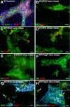
5-HT4 agonists promote neurogenesis in germinal niches in adult mouse gut. BrdU-labeled cells in a neuronal lineage were identified with antibodies to HuC/D. A, WT mouse, no exposure to 5-HT4 agonists, 2 weeks chase. No neurons (HuC/D) or glia (S100β) labeled with BrdU. DNA stained with bis. Scale bar, 20 μm. B, KO mouse, RS67506-treated, 2 weeks chase. No neurons (HuC/D) were labeled with BrdU. Scale bar, 16 μm. C, D, WT mice, exposed to 5-HT4 agonists, 2 weeks chase. Cells in clusters (germinal niches) adjacent to myenteric ganglia display coincident labeling with BrdU and punctate HuC/D. C, Ileum/RS67506. D, Colon/RS67506. Scale bar, 16 μm. E, F, WT mice, exposed to 5-HT4 agonists, 4 weeks chase. BrdU-labeled nuclei are present in extraganglionic cells with punctate HuC/D immunoreactivity (germinal niches). E, Ileum/RS67506. F, Colon/tegaserod. Scale bar, 16 μm. G, H, WT mice, colon, 5-HT4 agonists, 4 weeks chase. BrdU was incorporated into the nuclei of extraganglionic cells that were coimmunostained with the early neural marker DCX (arrows and arrowhead). Myenteric ganglia are identified by their immunostaining with antibodies to the neuronal marker, PGP9.5. The BrdU/DCX-colabeled cells are not in the same focal plane as the myenteric ganglia that are PGP9.5 immunoreactive. G, RS67506. H, Tegaserod. An extraganglionic DCX-immunoreactive cell that incorporated BrdU (arrowhead) is shown at higher magnification in the inset in H. Scale bars: G, H, 20 μm; H, inset, 6 μm.
Figure 8.
Figure 8.
The location of BrdU-labeled HuC/D-immunoreactive cells was studied by analyzing z-stacks of deconvoluted images through LMMP preparations of WT mouse colon at 4, 16, and 24 weeks of chase. 5-HT4 receptors were stimulated with RS67506 (A–C) or tegaserod (D). The immunoreactivity HuC/D (green) was used as a neuronal marker and that of BrdU (red) was used to mark cells undergoing mitosis during the pulse of BrdU. A, Four weeks of chase. Cells that are doubly labeled with antibodies to HuC/D and BrdU are still extraganglionic. The center panel shows a rotated image of the three-dimensional stack to provide a lateral view (1 unit = 8.2 μm); the panel at the left shows a focal plane imaged close to the level of the longitudinal muscle and the panel at the right illustrates a focal plane through the lower surface of a myenteric ganglion, which contains characteristic HuC/D-immunoreactive mature neurons that are not labeled with BrdU. The germinal niche is extraganglionic and located well below the ganglion. B–D, The diagrams at the left show the location of newly generated neuroblasts (cells that are colabeled with antibodies to BrdU and HuC/D; red hexagons), in relation to the longitudinal muscle (mauve ovals) and the nearest myenteric ganglion (green oval). The corresponding projection images from three-dimensional stack viewed vertically are illustrated to the right of the diagrams. B, Four weeks of chase. C, Sixteen weeks of chase. D, Twenty-four weeks of chase. Between 4 and 16 weeks, the newly generated neuroblasts moved closer to a ganglion, but newly generated neurons were not found in a ganglion until 24 weeks of chase. Scale bar, 6 μm.
Figure 9.
Figure 9.
Cells that display 5-HT4-induced BrdU incorporation express markers associated with crest-derived progenitors developing in a neuronal lineage. A–C, WT mouse treated with RS67506. After 4 weeks of chase, BrdU is not incorporated by cells that immunostained with antibodies to the glial markers B-FABP (A), GFAP (B), and S100β (C). Scale bar, 16 μm. D–F, WT mice, RS67506-treated. After 4 weeks of chase, BrdU is incorporated into extraganglionic Hu-immunoreactive cells that were labeled with antibodies to Sox10 (D, arrow), Phox2b (E1, arrow), or nestin (F1, arrow). Scale bar, 16 μm. The position of mature neurons in myenteric ganglia is shown by their HuC/D immunoreactivity. A typical myenteric ganglion is shown in E2 (Hu and Phox2b, myenteric neurons; Sox10, enteric glia). Note that mature neurons in C–F are intraganglionic and thus located in a focal plane that is different from that of the BrdU-immunoreactive extraganglionic germinal niche cells that coimmunostain with antibodies to HuC/D, Sox10 (D), Phox2b (E1), or nestin (F1, F2). G, In a WT mouse treated with RS67506, the cells that incorporate BrdU are located on a focal plane that is different from that of myenteric neurons labeled with HuC/D after 4 weeks of chase. Scale bar, 16 μm. H, WT mouse, RS67506-treated, 4 weeks chase. Cells that display 5-HT4-stimulated BrdU incorporation are Msi1 immunoreactive, which are adjacent to myenteric ganglia or near HuC/D-immunoreactive neurons. The Msi1-immunoreactive cell that incorporated BrdU appears to be acquiring perinuclear HuC/D immunoreactivity (arrow). This cell may be in transition from a stem or precursor cell to a progenitor in a neuronal lineage. Scale bar, 16 μm. I, Immunoblot. Msi1 immunoreactivity (39 kDa) can be detected not only in the intestinal mucosa and hippocampus but also in the intestinal LMMP. Antibodies to α-tubulin (55 kDa) were used as loading control. J, Presumed model to explain the extraganglionic location of BrdU-labeled cells that coexpress markers associated with crest-derived neural/glial precursors. Stem cells are located outside of ganglia, in which the terminal mitoses of neuronal precursors occur; progeny committed to a neuronal lineage then slowly migrate into ganglia. K, In-cell ELISA quantitation of BrdU incorporation into myenteric neurons isolated from LMMP of adult mice after 4 weeks of chase. Data are normalized to the vehicle controls for WT and KO mice (white bars). Exposure of WT mice to RS67506 or tegaserod significantly increased BrdU incorporation (**p < 0.01 vs WT). Neither RS67506 nor tegaserod increases BrdU incorporation in KO mice; tegaserod exposure reduced BrdU incorporation in the KO animals (**p < 0.01 vs KO). The number of mice per group was six. Error bars indicate SEM.
Figure 10.
Figure 10.
Cell proliferation is reduced and autophagy is increased in KO mice. A, B, Proliferating cells were detected with antibodies to Ki67 and neurons with antibodies to HuC/D. A, WT mice. Ki67 labeled nuclei of small extraganglionic clusters of cells with punctate HuC/D immunoreactivity (arrow). B, KO mice. No coincident labeling of cells with Ki67 and HuC/D is seen. Scale bar, 50 μm. C, D, Synaptotagmin- and peripherin-immunostained LMMP preparations isolated from WT (C) and KO (D, D′) mice. Peripherin-immunoreactive retraction bulbs (arrows) are found at the ends of neurites in circular muscle, tertiary plexus, and within myenteric ganglia in KO but not in WT mice. The degenerating terminals (retraction bulbs; arrows) extending from intact varicose axons are shown in D′. Many more retraction bulbs were found in LMMP from KO than WT mice. Scale bar, 50 μm. E, F, The autophagy marker, LC3B, is located immunocytochemically in HuD-expressing neurons and the neuropil of myenteric ganglia in WT (E) and KO (F) mice. Scale bar, 25 μm. G, Immunoblot showing LC3B immunoreactivity (16 kDa) extracted from membrane fractions from LMMP preparations of murine colons. The relative intensity of LC3B immunoreactivity was normalized to that of α-tubulin (55 kDa). The relative intensity of LC3B immunoreactivity of KO mice is significantly greater than that of WT mice (**p < 0.01; n = 3). The number of mice per group was three. Error bars indicate SEM. H, I, Electron micrographs of the myenteric plexus of WT (H) and KO (I) mice. cb, Nerve cell body; a, axon; v, varicosities; nu, nucleus. Autophagy is rare in the ENS of WT (H) but prominent in that of KO (I) mice. Autophagic structures (arrows) were found in neuronal cell bodies (not illustrated) and in dilated axonal varicosities (I, I′). Scale bar, 500 nm.

References

    1. Abràmoff MD, Magalhães PJ, Ram SJ. Image processing with ImageJ. Biophotonics Int. 2004;11:36–42.
    1. Almond S, Lindley RM, Kenny SE, Connell MG, Edgar DH. Characterization and transplantation of enteric nervous system progenitor cells. Gut. 2007;56:489–496. - PMC - PubMed
    1. Baetge G, Pintar JE, Gershon MD. Transiently catecholaminergic (TC) cells in the bowel of the fetal rat: precursors of noncatecholaminergic enteric neurons. Dev Biol. 1990;141:353–380. - PubMed
    1. Barami K, Iversen K, Furneaux H, Goldman SA. Hu protein as an early marker of neuronal phenotypic differentiation by subependymal zone cells of the adult songbird forebrain. J Neurobiol. 1995;28:82–101. - PubMed
    1. Beattie DT, Smith JA, Marquess D, Vickery RG, Armstrong SR, Pulido-Rios T, McCullough JL, Sandlund C, Richardson C, Mai N, Humphrey PP. The 5-HT4 receptor agonist, tegaserod, is a potent 5-HT2B receptor antagonist in vitro and in vivo. Br J Pharmacol. 2004;143:549–560. - PMC - PubMed

Publication types

MeSH terms

LinkOut - more resources