

doi: 10.1523/JNEUROSCI.1319-09.2009.
Boundary vector cells in the subiculum of the hippocampal formation
Affiliations
- PMID: 19657030
- PMCID: PMC2736390
- DOI: 10.1523/JNEUROSCI.1319-09.2009
Item in Clipboard
Boundary vector cells in the subiculum of the hippocampal formation
J Neurosci.
.
Abstract
"Boundary vector cells" were predicted to exist by computational models of the environmental inputs underlying the spatial firing patterns of hippocampal place cells (O'Keefe and Burgess, 1996; Burgess et al., 2000; Hartley et al., 2000). Here, we report the existence of cells fulfilling this description in recordings from the subiculum of freely moving rats. These cells may contribute environmental information to place cell firing, complementing path integrative information. Their relationship to other cell types, including medial entorhinal "border cells," is discussed.
Figures
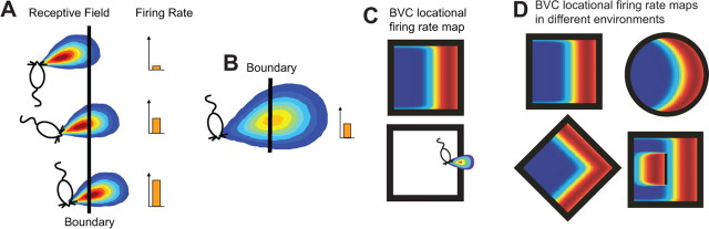
The BVC model. A BVC responds maximally when a boundary is perceived at a preferred distance and allocentric direction from the animal, regardless of the animal's heading direction. A, The receptive field of a BVC tuned to respond to a barrier at a short distance east-northeast from the animal. B, BVCs tuned to respond to barriers farther from the animal will have broader receptive fields. C, The firing field (firing rate as a function of the animal's location; top) for a BVC with a receptive field tuned to respond to a boundary at a short distance to the east (bottom). D, Predicted firing fields in different environments for the BVC shown in C. Insertion of a barrier causes a doubling of the field (bottom right panel). Figures are adapted from Hartley et al. (2000) and Barry and Burgess (2007).
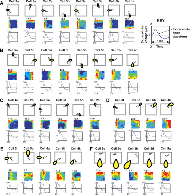
Putative BVCs recorded from dorsal subiculum. Firing fields (middle rows) from trials in environment a and corresponding BVC receptive fields (top rows). Cells are identified by animal number, followed by cell letter. The number shown top left of the firing rate map denotes peak rate in hertz after smoothing [firing rate map methods as in the study by Wills et al. (2005)]. The bottom rows show the averaged waveform taken from the tetrode channel with the largest peak-to-trough amplitude: y-axis is extracellular voltage (in microvolts, negative upward); x-axis is time (1 ms).
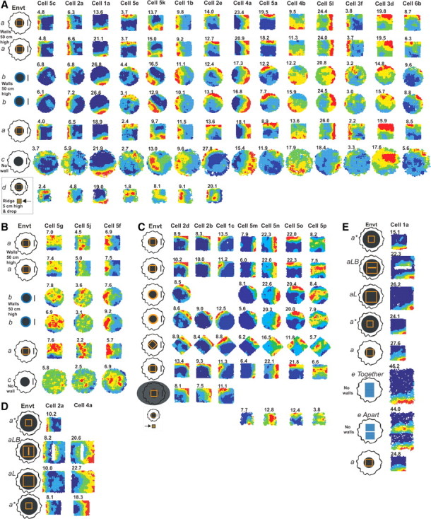
BVC firing fields in different environments. A, Firing fields of 14 subicular BVCs in different environments a–d (see supplemental material, available at www.jneurosci.org , for a full description). Environment (Envt) a, 62 × 62 × 50-cm-high beige square box made of morph material; Envt b, 79-cm-diameter, circular-walled, wooden light-gray enclosure; Envt c, the 90-cm-diameter floor of Envts a and b; Envt d, 39-cm-sided, square holding platform with 5-cm-high ridges located 2 m south of Envt a. B, Firing fields of three BVCs in environments a–c, which have more diffuse firing in Envt c than those shown in A. C, Firing fields of seven BVCs tested in square-, circular-, and diamond- shaped environments (all made of the same morph box material). Three of these were also recorded in complete darkness (row 7), and four were recorded on the holding platform (Envt d, row 8). D, E, Cells 2a and 4a (D) and cell 1a (E) under additional environmental manipulations. Envt a* is Envt a placed on a larger platform; Envts aL and aLB are larger square environments made of the same morph material (aLB also contains a barrier). In the e Apart condition, a 13 cm gap is interposed between the two elevated, rectangular “drop” platforms of the e Together condition. Cells 2a and 5c are adapted from Barry et al. (2006).
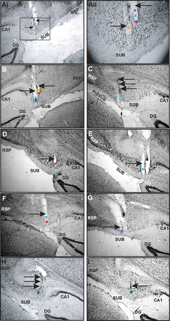
Recording locations of BVCs. Nissl-stained sections of the dorsal subiculum are shown. Colored squares indicate estimated locations of BVCs; arrows indicate tracks of recording tetrodes. Cells recorded at depths 50 μm apart are shown at the same location. Ai, Tetrode tracks in subiculum of rat 5. Aii, Close-up of the rectangular area indicated in Ai. Orange square, Estimated location of cell 5j; purple square, cell 5h; blue square, cell 5g; red square, cell 5m-n. B, Purple square, cell 5o-p; green square, cells 5a, 5e, 5f, and 5k; blue square, cell 5d; red square, cell 5c; orange square, cells 5l and 5b; yellow square, cell 5i. C, Purple square, cell 6a; blue square, cell 6b. D, Red square, cell 1a; blue square, cell 1c. E, Blue square, cell 1b. F, Red square, cells 2a and 2e; blue square, cell 2c. G, Blue square, cell 2b; purple square, cell 2d. H, Green square, cells 3a, 3e, and 3g; blue square, cell 3d. The estimated location of cell 3h is similar to cells 3a, 3e, and 3g, but 90 μm posterior (data not shown). I, Green square, cell 3b-c; blue square, cell 3f. SUB, Subiculum; RSP, retrosplenial cortex; DG, dentate gyrus.
Comment in
-
Are the boundary-related cells in the subiculum boundary-vector cells?J Neurosci. 2009 Oct 28;29(43):13429-31. doi: 10.1523/JNEUROSCI.4176-09.2009. J Neurosci. 2009. PMID: 19864554 Free PMC article. No abstract available.
References
Publication types
MeSH terms
Grants and funding
- 082507/WT_/Wellcome Trust/United Kingdom
- G01342X/1/BB_/Biotechnology and Biological Sciences Research Council/United Kingdom
- G0501672(76328)/MRC_/Medical Research Council/United Kingdom
- BB/G01342X/1/BB_/Biotechnology and Biological Sciences Research Council/United Kingdom
- G0501672/MRC_/Medical Research Council/United Kingdom
LinkOut - more resources
Full Text Sources
Other Literature Sources
Molecular Biology Databases