. 2009 Aug 5;29(31):9794-808.
doi: 10.1523/JNEUROSCI.1544-09.2009.

Transient receptor potential canonical 5 channels activate Ca2+/calmodulin kinase Igamma to promote axon formation in hippocampal neurons

Affiliations

Transient receptor potential canonical 5 channels activate Ca2+/calmodulin kinase Igamma to promote axon formation in hippocampal neurons

Monika A Davare et al. J Neurosci. .

Abstract

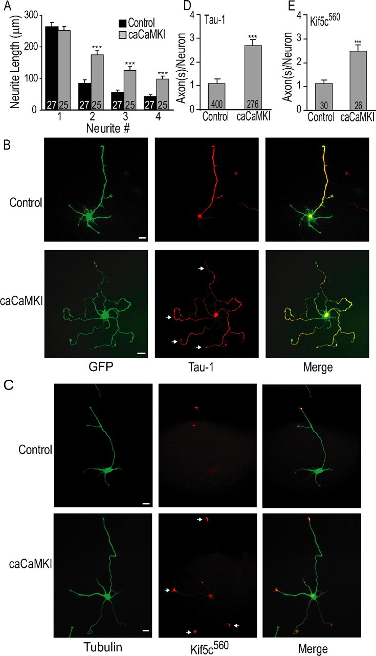
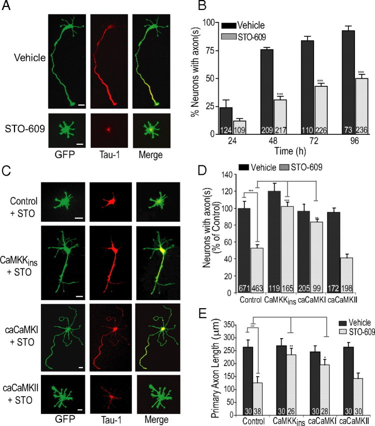
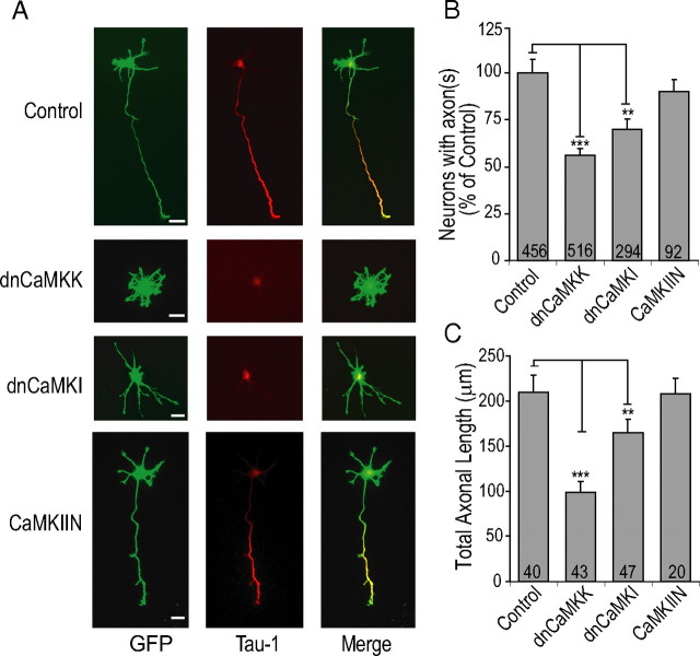
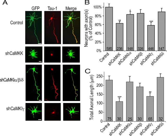
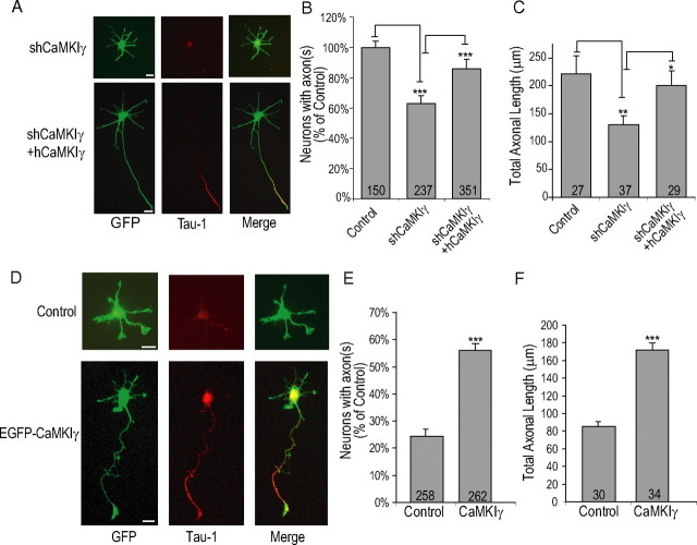
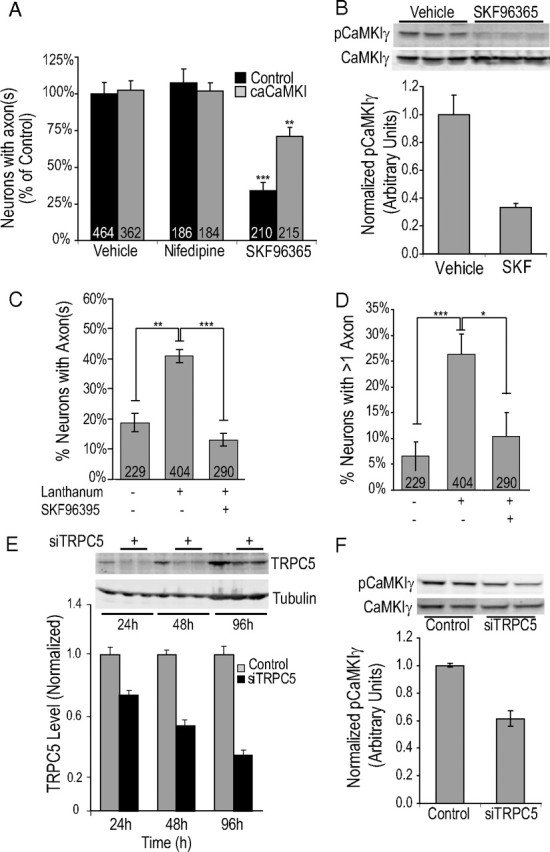
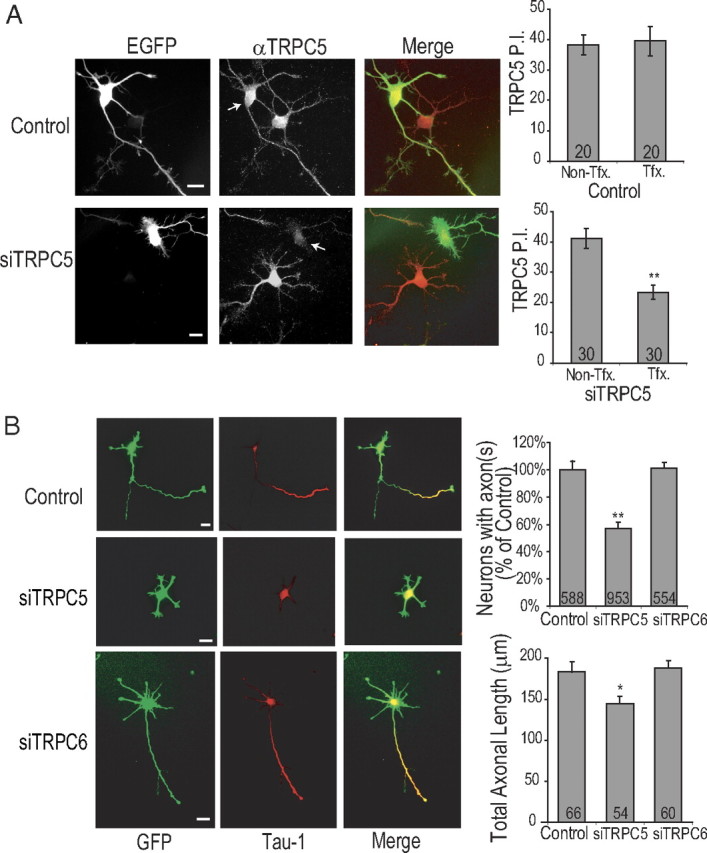
Functionality of neurons is dependent on their compartmentalized polarization of dendrites and an axon. The rapid and selective outgrowth of one neurite, relative to the others, to form the axon is critical in initiating neuronal polarity. Axonogenesis is regulated in part by an optimal intracellular calcium concentration. Our investigation of Ca(2+)-signaling pathways involved in axon formation using cultured hippocampal neurons demonstrates a role for Ca(2+)/calmodulin kinase kinase (CaMKK) and its downstream target Ca(2+)/calmodulin kinase I (CaMKI). Expression of constitutively active CaMKI induced formation of multiple axons, whereas blocking CaMKK or CaMKI activity with pharmacological, dominant-negative, or short hairpin RNA (shRNA) methods significantly inhibited axon formation. CaMKK signals via the gamma-isoform of CaMKI as shRNA to CaMKIgamma, but not the other CaMKI isoforms, inhibited axon formation. Furthermore, overexpression of wild-type CaMKIgamma, but not a mutant incapable of membrane association, accelerated the rate of axon formation. Pharmacological or small interfering RNA inhibition of transient receptor potential canonical 5 (TRPC5) channels, which are present in developing axonal growth cones, suppressed CaMKK-mediated activation of CaMKIgamma as well as axon formation. We demonstrate using biochemical fractionation and immunocytochemistry that CaMKIgamma and TRPC5 colocalize to lipid rafts. These results are consistent with a model in which highly localized calcium influx through the TRPC5 channels activates CaMKK and CaMKIgamma, which subsequently promote axon formation.


Figures

Figure 1.
Figure 1.
Constitutively active CaMKI induces multiple axon formation in hippocampal neurons. A, Average neurite length at 48 h from neurons transfected either with control plasmid (empty vector) or caCaMKI. B, Low-density E18 hippocampal cultures were electroporated before plating with either pCAGGS (control; top panel) or pCAGGS-caCaMKI (bottom panel) and soluble EGFP (marker for transfection) for 48 h followed by fixation and staining as described in Materials and Methods. Representative immunofluorescent images demonstrate formation of single Tau-1-positive neurite defined as the axon in control cells. In contrast, expression of caCaMKI strongly increases the number of Tau-1-positive axons. C, GFP-tagged Kif5C560 (pseudocolored red) was cotransfected with control or caCaMKI before plating, and neurons were fixed and stained with anti-β-tubulin (pseudocolored green) at 48 h. Representative images are shown. D, E, Quantification (means ± SEM) of number of Tau-1-positive axons per neuron from three independent experiments (D) and GFP-tagged Kif5C560-containing neurite tips from control and caCaMKI (E). Scale bars: 20 μm (all panels). Here and in all subsequent figures, all graphs show means ± SEM (n values are shown as numerical insets within bars of graphs; where indicated, p ≤ 0.05, **p ≤ 0.005, ***p ≤ 0.0005 by Student's t test).
Figure 2.
Figure 2.
Inhibition of CaMKK by STO-609 chronically suppresses axon formation. A, Low-density hippocampal cultures electroporated before plating with soluble EGFP were incubated with vehicle (control) or 5 μm STO-609 from 2 h until fixation at 48 h. Representative images with axonal marker Tau-1 shows normal formation of a single axon in control neurons with suppressed axon formation in neurons incubated in STO-609. B, STO-609 time course with neurons fixed at 24, 48, 72, and 96 h reveals chronic partial block of axon formation. C–E, Expression of STO-609-insensitive CaMKK (CaMKKins) or caCaMKI, but not caCaMKII, rescue STO-609 inhibition of axon formation and outgrowth. All inhibitor and inhibitor rescue data are shown normalized to control conditions set to 100% [60–90% of neurons polarized under basal (control) conditions] in this and subsequent figures. Representative images from STO-609-insensitive CaMKK, caCaMKI, and caCaMKII are shown in C. Quantification of three independent experiments are shown in D. Average axonal length as measured by length of primary Tau-1-positive axon is shown in graph in E. Scale bars: 20 μm (all panels).
Figure 3.
Figure 3.
dnCaMKK and dnCaMKI inhibit axon formation and decrease axonal length. A, E18 hippocampal neurons were coelectroporated with control (empty vector), dnCaMKK, dnCaMKI, or CaMKIIN containing plasmid and soluble EGFP before plating. A shows representative images of neurons fixed at 48 h and stained with the axonal marker Tau-1. B, Quantification of axon formation as measured by number of neurons with Tau-1-positive projections from three to four independent experiments. C, Quantification of average axonal length as measured by the length of the primary Tau-1-positive neurite. Scale bars: 20 μm (all panels).
Figure 4.
Figure 4.
Knockdown of CaMKK and CaMKIγ protein expression inhibits axonogenesis and decreases axonal length. A, E18 hippocampal neurons were electroporated before plating with pMU6pro alone (control) or pMU6pro containing shCaMKK, shCaMKIα, shCaMKIβ, shCaMKIδ, or shCaMKIγ. Between 2 and 48 h in culture, low-density cultures of electroporated neurons were incubated with 20 μm SP600125 to suppress intrinsic neuronal polarization to achieve effective knockdown with the plasmid based shRNA (see Results). At 48–72 h in culture, electroporated neurons were released from SP600125 block by transfer of coverslips to new glial culture plates for an additional 48 h. Representative images of neurons fixed and stained with Tau-1 at 96 h are shown. The third from top panel shows staining from CaMKIα shRNA. This image is also representative of neurons transfected with shCaMKIβ or shCaMKIδ. B, Summary of three to five independent experiments. C, Average axonal length as measured by length of primary process containing Tau-1 staining from three independent experiments. Scale bars: 20 μm (all panels).
Figure 5.
Figure 5.
CaMKIγ expression promotes axon formation and increases axonal length. A–C, Coexpression of shRNA-insensitive human CaMKIγ rescues the effect of shCaMKIγ. Representative images are shown in A. Quantification of percentage of neurons exhibiting an axon and total axonal length from shCaMKIγ and shCaMKIγ plus rescue plasmid containing hCaMKIγ are shown in B and C, respectively. D, Representative images of EGFP-wtCaMKIγ induced accelerated axon formation at 24 h in culture. Controls contained soluble EGFP. E, F, Quantification of percentage of neurons exhibiting an axon at 24 h and total axonal length of neurons polarized at 24 h, respectively. Scale bars: 20 μm (all panels).
Figure 6.
Figure 6.
Regulation of CaMKIγ and axon formation by TRPC channel. A, Low-density hippocampal neurons electroporated before plating were incubated for 46 h (2 h after plating until fixation at 48 h) in culture with vehicle, the L-type channel blocker nifedipine (10 μm), or the TRPC blocker SKF96395 (3 μm). Neurons fixed at 48 h were stained with the axonal marker Tau-1. Quantification of results from three independent experiments is shown. B, Representative blot showing regulation of CaMKIγ by TRPC antagonist, SKF96365. P0 hippocampal neurons were electroporated with Flag-CaMKIγ before plating and 24 h later exposed to 50 μm SKF96365 for 60 min. Lysates were immunoblotted with anti-phospho-CaMKI antibody (top blot) or anti-Flag antibody to detect total CaMKIγ (bottom blot). The bottom graph shows average normalized phospho-CaMKIγ. C, D, Graphs depict quantification of axon formation (C) and induction of multiple axons (D) in E18 neurons that were incubated for 20 h (from 2 to 22 h) with growth medium containing 100 μm lanthanum chloride, a selective activator of TRPC4 and -5 channels. E, High-density hippocampal neurons were electroporated with 2.5 μg of siTRPC5 and harvested at the indicated times with SDS sample buffer. Western blots show the time course of reduction of endogenous TRPC5 (top blot) and β-tubulin (bottom blot) with siTRPC5. The bottom graph shows quantification of TRPC5 levels relative to tubulin at 24, 48, 72 h (with control at 24 h set to arbitrary unit of 1). F, Flag-CaMKIγ and control siRNA or siTRPC5 were coelectroporated in P0 hippocampal neurons and harvested after 48–72 h in SDS sample buffer. Phospho-CaMKIγ and total CaMKIγ (Flag) were detected by multiplexed Western blotting and the bottom graph represents summary of ratio of pCaMKIγ/total CaMKIγ from four independent experiments.
Figure 7.
Figure 7.
Regulation of axon formation by TRPC5 channels. A, E18 Hippocampal neurons were electroporated before plating with control siRNA or siTRPC5 plus soluble EGFP. Neurons were fixed at 48 h after electroporation and stained with anti-TRPC5 polyclonal antibody and imaged as described in Materials and Methods. Representative images from control siRNA (top) and siTRPC5 (bottom) are shown in left panel and the graphs summarizing quantification (y-axis shows TRPC5 P.I. in arbitrary units) are shown on right. B, E18 neurons were electroporated before plating with control siRNA, siTRPC5, or siTRPC6. Neurons were fixed at 48 h and evaluated for axon formation by staining with anti-Tau-1 antibody (see Materials and Methods). Representative images are shown in left panel(s) and quantification of percentage of polarized neurons (top graph) and length of (bottom graph) in the neurons that did polarize is shown on right. Scale bars: 20 μm (all panels).
Figure 8.
Figure 8.
CaMKIγ and TRPC5 colocalize with lipid raft markers in rat brain and cultured hippocampal neurons. A, Endogenous CaMKK, CaMKIγ, and TRPC5 are present in the lipid raft fraction (fractions 4) as confirmed by enrichment of the raft marker Flotillin-1 in fraction 4 of a discontinuous sucrose density gradient (fractions 1–4, 5% sucrose; fractions 5–11, 35% sucrose; fraction 12, 50% sucrose) from P0 rat forebrains. The input is the crude homogenate before any centrifugation steps separating cytosol and membranes. B, Immunofluorescent staining of endogenous TRPC5 (pseudocolored red) and endogenous Flotillin-1 (pseudocolored green) in axonal growth cone of E18 hippocampal neuron. Scale bar: 5 μm. C, Colocalization of Alexa Fluor 546-CTxB labeled raft marker GM1 (as described in Materials and Methods) and EGFP-CaMKIγ in DIV 1 hippocampal neurons. Scale bar: D, 20 μm. Immunofluorescent staining of endogenous β-tubulin, TRPC5 with electroporated EGFP-CaMKIγ in stage 2 (top row) and stage 3 (bottom row) neurons shows extensive colocalization of TRPC5 and EGFP-CaMKIγ. In final column, the images are a merge of all three channels, with β-tubulin pseudocolored blue, EGFP-CaMKIγ pseudocolored green, and endogenous TRPC5 pseudocolored red in stage 2 and stage 3 hippocampal neurons. The insets show additional magnification of largest growth cones. Scale bars: β-tubulin (bottom) panel, 10 μm; EGFP-CaMKIγ (bottom inset showing growth cone), 5 μm. E, Quantification of neurons with Tau-1-positive axons expressing wild-type CaMKIγ, CaMKIγDPP (mutant lacking prenylation and palmitoylation sites), or CaMKIα fixed at 24 h to evaluate acceleration of axonogenesis. F, Quantification of Tau-1-positive axons in neurons expressing wild-type CaMKIγ alone or wild-type CaMKIγ plus siTRPC5 at 24 h. Note, in D and E, neurons are fixed at 24 h to test mutant CaMKIγ and siTRPC5 in acceleration of axon formation assay. G, Quantification of rescue experiment from neurons expressing control, siTRPC5, or siTRPC5 plus rescue with caCaMKIγ.

Comment in

References

    1. Alfonso S, Benito O, Alicia S, Angélica Z, Patricia G, Diana K, Vaca L, Luis V. Regulation of the cellular localization and function of human transient receptor potential channel 1 by other members of the TRPC family. Cell Calcium. 2008;43:375–387. - PubMed
    1. Alicia S, Angélica Z, Carlos S, Alfonso S, Vaca L. STIM1 converts TRPC1 from a receptor-operated to a store-operated channel: moving TRPC1 in and out of lipid rafts. Cell Calcium. 2008;44:479–491. - PubMed
    1. Ambudkar IS. Ca2+ signaling microdomains: platforms for the assembly and regulation of TRPC channels. Trends Pharmacol Sci. 2006;27:25–32. - PubMed
    1. Ambudkar IS, Ong HL. Organization and function of TRPC channelosomes. Pflugers Arch. 2007;455:187–200. - PubMed
    1. Ambudkar IS, Bandyopadhyay BC, Liu X, Lockwich TP, Paria B, Ong HL. Functional organization of TRPC-Ca2+ channels and regulation of calcium microdomains. Cell Calcium. 2006;40:495–504. - PubMed

Publication types

MeSH terms

Substances