

Local uteroplacental influences are responsible for the induction of uterine artery myogenic tone during rat pregnancy
- PMID: 19657140
- PMCID: PMC2759862
- DOI: 10.1177/1933719109340927
Local uteroplacental influences are responsible for the induction of uterine artery myogenic tone during rat pregnancy
Abstract
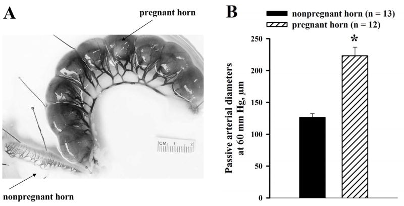
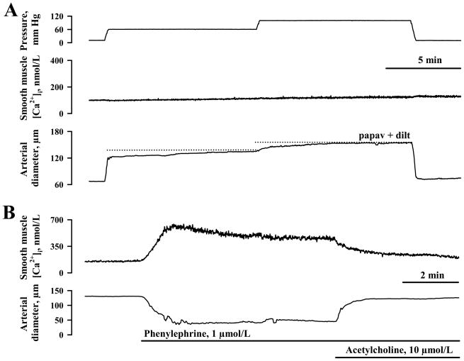
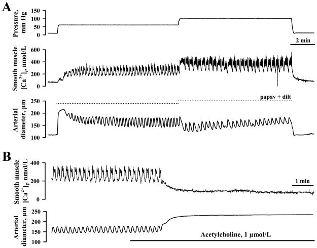
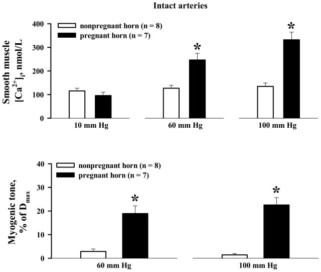
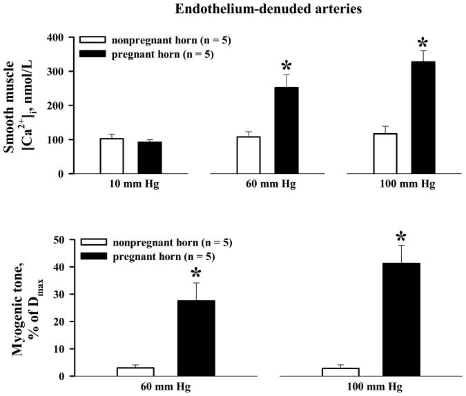
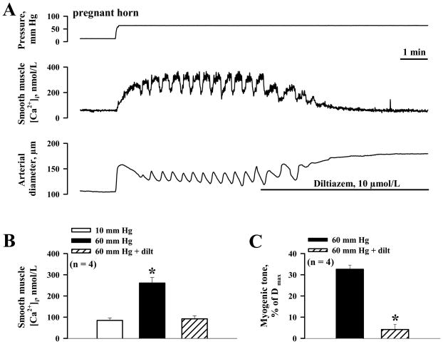
Uterine artery constrictor responses to elevation of intraluminal pressure (myogenic tone) are considerably enhanced in late pregnant rats, although the underlying causes remain unknown. A single uterine horn ligation model was used to differentiate local from systemic influences, and to test the hypothesis that factors associated with the site of placentation, rather than systemic hormonal changes, are primarily involved in the induction of this adaptive process. Radial uterine arteries were dissected from the gravid and nongravid uterine horns of late pregnant rats, cannulated, and pressurized. Changes in arterial diameter and smooth muscle [Ca(2+)](i) in response to the elevation of intraluminal pressure were studied using intact and endothelium-denuded arteries loaded with the ratiometric Ca(2+)-sensitive dye fura-2. Elevations of pressure from 10 to 60 and 100 mm Hg resulted in passive arterial distention of arteries from nongravid horns with a minor change in [Ca(2+)](i). In contrast, arteries from gravid horns developed myogenic tone associated with a significant elevation in [Ca(2+)](i). Synchronous oscillations in [Ca(2+)](i) and lumen diameter were frequently observed in vessels from gravid horns. Endothelial denudation augmented tone in the gravid horn but did not uncover myogenic tone in vessels from the nongravid horn. In summary, pregnancy-associated uterine artery myogenic behavior is due to an upregulation of calcium-handling mechanisms, occurs independently of the endothelium, and is induced by local uteroplacental influences.
Figures
Similar articles
-
Pregnancy Increases Ca2+ Sparks/Spontaneous Transient Outward Currents and Reduces Uterine Arterial Myogenic Tone.Hypertension. 2019 Mar;73(3):691-702. doi: 10.1161/HYPERTENSIONAHA.118.12484. Hypertension. 2019. PMID: 30661479 Free PMC article.
-
Increased uterine arterial tone, stiffness and remodeling with augmented matrix metalloproteinase-1 and -7 in uteroplacental ischemia-induced hypertensive pregnancy.Biochem Pharmacol. 2024 Oct;228:116227. doi: 10.1016/j.bcp.2024.116227. Epub 2024 Apr 19. Biochem Pharmacol. 2024. PMID: 38643908
-
Role of impaired endothelial cell Ca(2+) signaling in uteroplacental vascular dysfunction during diabetic rat pregnancy.Am J Physiol Heart Circ Physiol. 2013 Apr 1;304(7):H935-45. doi: 10.1152/ajpheart.00513.2012. Epub 2013 Feb 1. Am J Physiol Heart Circ Physiol. 2013. PMID: 23376827 Free PMC article.
-
Decreased function of voltage-gated potassium channels contributes to augmented myogenic tone of uterine arteries in late pregnancy.Am J Physiol Heart Circ Physiol. 2008 Jan;294(1):H272-84. doi: 10.1152/ajpheart.00216.2007. Epub 2007 Nov 2. Am J Physiol Heart Circ Physiol. 2008. PMID: 17982020
-
Effects of the ovary and conceptus on uterine blood flow in the pig.J Reprod Fertil Suppl. 1985;33:83-90. J Reprod Fertil Suppl. 1985. PMID: 2418203 Review.
Cited by
-
Enhanced Vascular Smooth Muscle Calcium Sensitivity and Loss of Endothelial Vasodilator Influence Contribute to Myogenic Tone Development in Rat Radial Uterine Arteries during Gestation.J Vasc Res. 2020;57(3):126-135. doi: 10.1159/000505670. Epub 2020 Feb 27. J Vasc Res. 2020. PMID: 32106116 Free PMC article.
-
Uterine Vascular Control Preconception and During Pregnancy.Compr Physiol. 2021 Jun 1;11(3):1871-1893. doi: 10.1002/cphy.c190015. Compr Physiol. 2021. PMID: 34061977 Free PMC article.
-
Pregnancy Increases Ca2+ Sparks/Spontaneous Transient Outward Currents and Reduces Uterine Arterial Myogenic Tone.Hypertension. 2019 Mar;73(3):691-702. doi: 10.1161/HYPERTENSIONAHA.118.12484. Hypertension. 2019. PMID: 30661479 Free PMC article.
-
Plasticity of the Maternal Vasculature During Pregnancy.Annu Rev Physiol. 2019 Feb 10;81:89-111. doi: 10.1146/annurev-physiol-020518-114435. Annu Rev Physiol. 2019. PMID: 30742784 Free PMC article. Review.
-
Effect of Oxidative Stress on the Estrogen-NOS-NO-KCa Channel Pathway in Uteroplacental Dysfunction: Its Implication in Pregnancy Complications.Oxid Med Cell Longev. 2019 Feb 10;2019:9194269. doi: 10.1155/2019/9194269. eCollection 2019. Oxid Med Cell Longev. 2019. PMID: 30881600 Free PMC article. Review.
References
-
- Poston L, McCarthy AL, Ritter JM. Control of vascular resistance in the maternal and feto-placental arterial beds. Pharmacol Ther. 1995;65(2):215–239. - PubMed
-
- Sladek SM, Magness RR, Conrad KP. Nitric oxide and pregnancy. Am J PhysiolRegul Integr Comp Physiol. 1997;272(2 pt 2):R441–R463. - PubMed
-
- Moll W. Structure adaptation and blood flow control in the uterine arterial system after hemochorial placentation. Eur J Obstet Gynecol Reprod Biol. 2003;110 (Suppl 1):S19–S27. - PubMed
-
- Magness RR, Rosenfeld CR, Hassan A, Shaul PW. Endothelial vasodilator production by uterine and systemic arteries. I. Effects of ANG II on [PGI]2 and NO in pregnancy. Am J Physiol Heart and Circ Physiol. 1996;270(6 pt 2):H1914–H1923. - PubMed
-
- Bird IM, Zhang L, Magness RR. Possible mechanisms underlying pregnancy-induced changes in uterine artery endothelial function. Am J Physiol Regul Integr Comp Physiol. 2003;284(2):R245–R258. - PubMed
Publication types
MeSH terms
Substances
Grants and funding
LinkOut - more resources
Full Text Sources
Miscellaneous
