. 2009 Aug 27;460(7259):1127-31.
doi: 10.1038/nature08248. Epub 2009 Aug 5.

L1 retrotransposition in human neural progenitor cells

Affiliations

L1 retrotransposition in human neural progenitor cells

Nicole G Coufal et al. Nature. .

Abstract

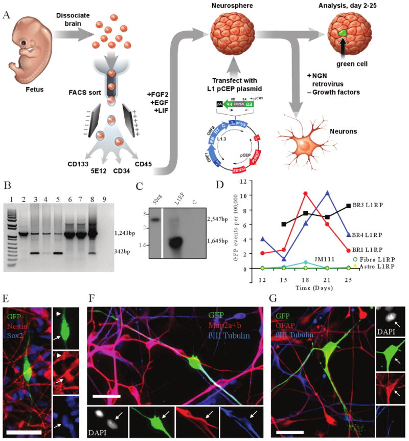
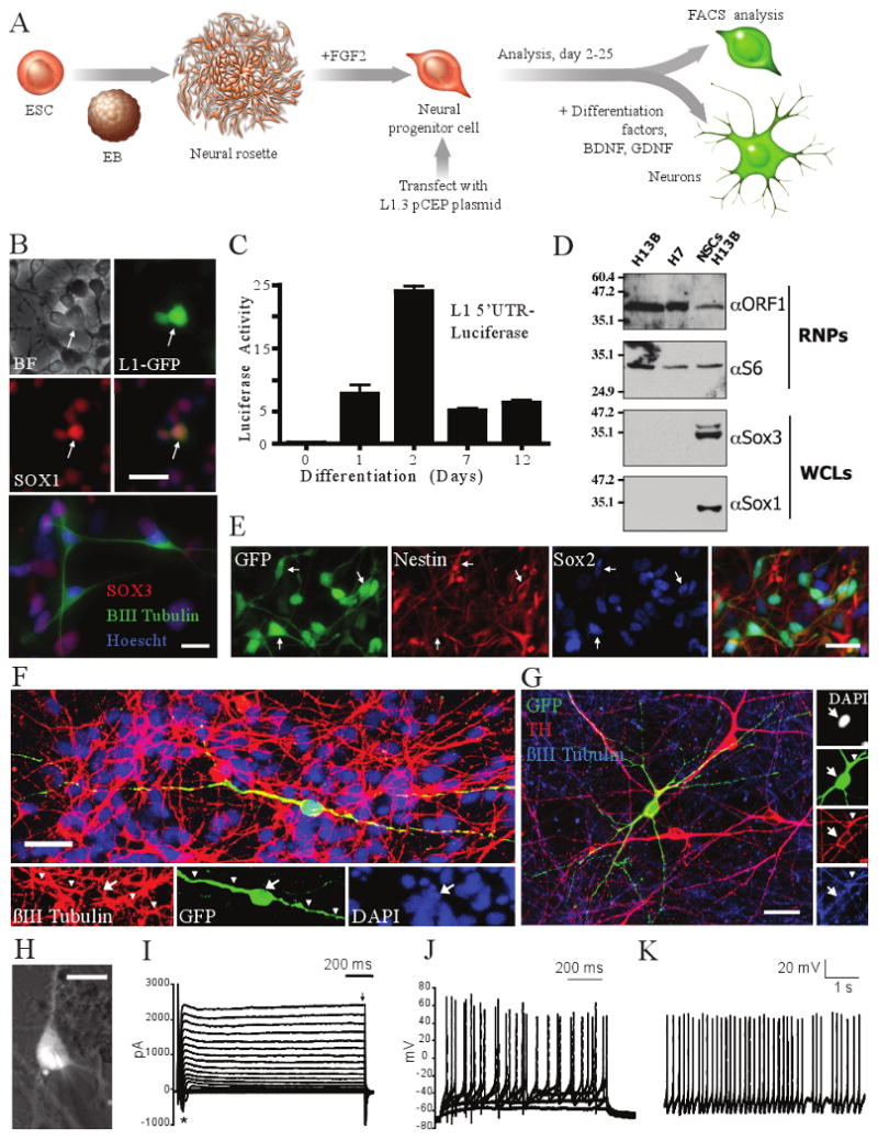
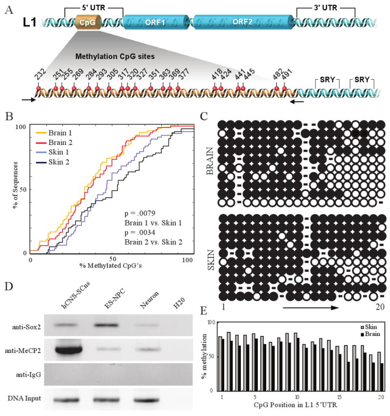
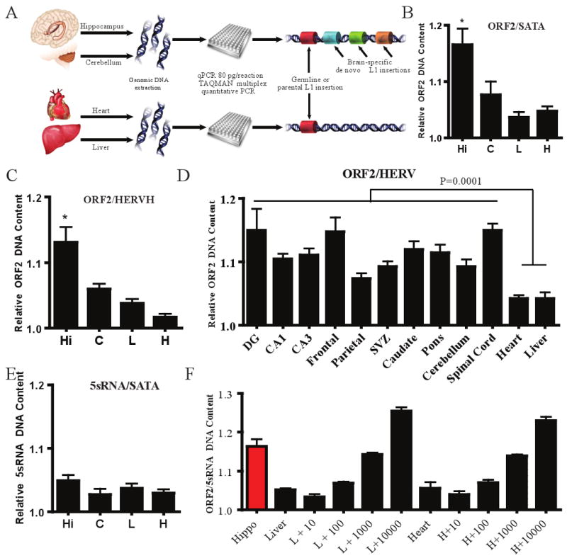
Long interspersed element 1 (LINE-1 or L1) retrotransposons have markedly affected the human genome. L1s must retrotranspose in the germ line or during early development to ensure their evolutionary success, yet the extent to which this process affects somatic cells is poorly understood. We previously demonstrated that engineered human L1s can retrotranspose in adult rat hippocampus progenitor cells in vitro and in the mouse brain in vivo. Here we demonstrate that neural progenitor cells isolated from human fetal brain and derived from human embryonic stem cells support the retrotransposition of engineered human L1s in vitro. Furthermore, we developed a quantitative multiplex polymerase chain reaction that detected an increase in the copy number of endogenous L1s in the hippocampus, and in several regions of adult human brains, when compared to the copy number of endogenous L1s in heart or liver genomic DNAs from the same donor. These data suggest that de novo L1 retrotransposition events may occur in the human brain and, in principle, have the potential to contribute to individual somatic mosaicism.


Figures

Figure 1
Figure 1. L1 retrotransposition in hCNS-SCns
a, Experimental rationale. b, PCR of genomic DNA; 1,243bp product contains the intron; 342bp product indicates intron loss and retrotransposition. Lane 1, weight standards; Lane 2, hCNS-SCns transfected with JM111/L1RP; Lanes 3-5, three hCNS-SCns lines transfected with L1RP, Lane 6-7, primary astrocytes and fibroblasts transfected with L1RP; Lane 8, positive control; Lane 9, water. c, Southern blot of hCNS-SCns (line FBR-BR3); 2,547bp band represents plasmid; 1,645bp band is diagnostic for genomic insertion. d, Time course of L1 retrotransposition. e, EGFP-positive cells express Nestin and Sox2. f, EGFP-positive cells can differentiate to neurons (βIII tubulin and Map2a+2b positive). g, EGFP-positive cells can differentiate into glia (GFAP positive, βIII tubulin negative). Scale bar = 25 μm, arrows indicate co-labeled cell body; arrowheads indicate co-labeled processes.
Figure 2
Figure 2. L1 retrotransposition in hESC-derived NPCs
a, Experimental rationale. b, L1 retrotransposition in H13B (top, LRE3-GFP) and H7 (bottom, LRE3-mneo1)-derived NPCs (BF = brightfield). G418-resistant foci can express progenitor (SOX3) and neuronal (βIII tubulin) markers. c, L1 5′ UTR is induced upon differentiation. d, H13B-derived NPCs express endogenous ORF1p. RNP= ribonucleoprotein particle samples; WCL= whole cell lysate. e-g, EGFP-positive, HUES6-derived NPCs express SOX2 and Nestin and can differentiate to be tyrosine hydroxylase (TH) positive. Scale bar = 25 μm. h, LRE-EGFP positive neuron from which (I)-(K) were obtained. Scale bar = 10 μm. i, Transient Na+ (asterisk) and sustained K+ currents (arrow) in response to voltage step depolarizations j, Suprathreshold responses to somatic current injections. k, Spontaneous action potentials (Vm = −50 mV). Arrows indicate cell soma co-localization; arrowheads indicate co-labeled processes.
Figure 3
Figure 3. Methylation analysis and chromatin immunoprecipitation (ChIP) for the endogenous human L1 5′ UTR
a, Schematic illustrating the L1 CpG island, and SRY/SOX2 binding sites. b, Cumulative distribution function (CDF) plot, comparing overall methylation and collapsing CpG sites into a single data point, (two-sample Kolmogorov-Smirnov test). c, Individual methylation of sequences exhibiting highest sequence similarity to consensus RC-L1s. Open circles = unmethylated, closed circles = methylated CpG dinucleotides. d, ChIP identifying MeCP2 and Sox2 occupying the endogenous human L1 promoter, extracts were analyzed by PCR towards the L1 5′ UTR SRY binding region (Sox2 immunoprecipitation) or CpG island region (MeCP2 immunoprecipitation). e, CpG dinucleotides exhibited higher methylation at the 5′ end of the CpG island; higher methylation overall was observed in skin samples.
Figure 4
Figure 4. Multiplex quantitative PCR analyses of L1 copy number in human tissues
a, Experimental schematic. b-c, Relative quantity of L1, standardized such that the lowest liver value was normalized to 1.0. Hi = hippocampus, C = cerebellum, H = heart, and L = liver. Additional L1 ORF2 assays with other internals controls, Fig. S9-10. Error bars all s.e.m. *, p<0.05 (repeated measures one-way ANOVA with Bonferroni correction, n=3 individuals, with 3 repeat samples from each tissue). d, Ten samples from various brain regions (n=3 individuals) compared to somatic liver and heart. One-way t-test, p ≤ 0.0001 with 34 degrees of freedom. e, Multiplexing of 5S rDNA with α -satellite indicated no significant change, p≤0.5054. f, Hippocampal tissue compared to liver and heart spiked with estimated plasmid copy numbers of L1 (10, 100, 1,000, and 10,000 copies).

Comment in

Similar articles

Cited by

References

    1. Muotri AR, et al. Somatic mosaicism in neuronal precursor cells mediated by L1 retrotransposition. Nature. 2005;435(7044):903–910. - PubMed
    1. Tang Y, Nyengaard JR, De Groot DM, Gundersen HJ. Synapse. 3. Vol. 41. New York, N.Y: 2001. Total regional and global number of synapses in the human brain neocortex; pp. 258–273. - PubMed
    1. Uchida N, et al. Direct isolation of human central nervous system stem cells. Proc Natl Acad Sci U S A. 2000;97(26):14720–14725. - PMC - PubMed
    1. Brouha B, et al. Hot L1s account for the bulk of retrotransposition in the human population. Proc Natl Acad Sci U S A. 2003;100(9):5280–5285. - PMC - PubMed
    1. Moran JV, et al. High frequency retrotransposition in cultured mammalian cells. Cell. 1996;87(5):917–927. - PubMed

Publication types