. 2009 Oct;12(10):1238-47.
doi: 10.1038/nn.2387. Epub 2009 Aug 5.

SOX6 controls dorsal progenitor identity and interneuron diversity during neocortical development

Affiliations

SOX6 controls dorsal progenitor identity and interneuron diversity during neocortical development

Eiman Azim et al. Nat Neurosci. 2009 Oct.

Abstract

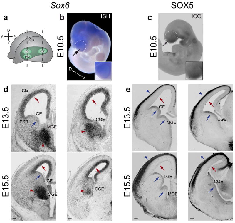
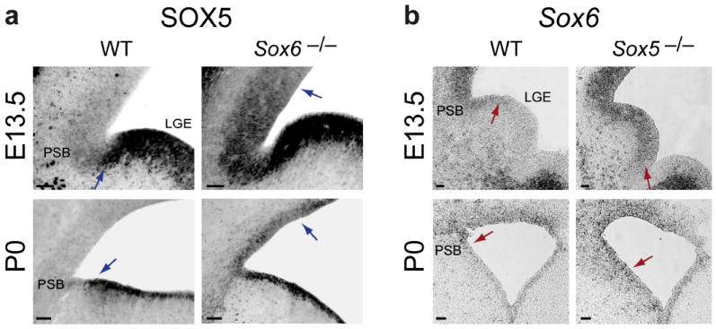
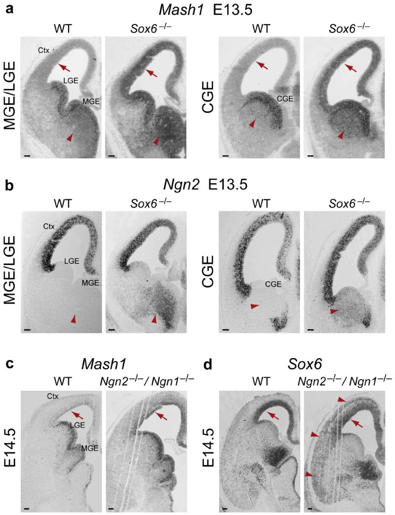
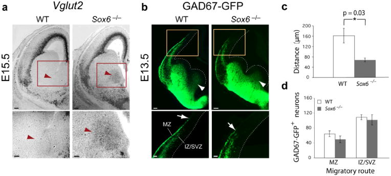
The neuronal diversity of the CNS emerges largely from controlled spatial and temporal segregation of cell type-specific molecular regulators. We found that the transcription factor SOX6 controls the molecular segregation of dorsal (pallial) from ventral (subpallial) telencephalic progenitors and the differentiation of cortical interneurons, regulating forebrain progenitor and interneuron heterogeneity. During corticogenesis in mice, SOX6 and SOX5 were largely mutually exclusively expressed in pallial and subpallial progenitors, respectively, and remained mutually exclusive in a reverse pattern in postmitotic neuronal progeny. Loss of SOX6 from pallial progenitors caused their inappropriate expression of normally subpallium-restricted developmental controls, conferring mixed dorsal-ventral identity. In postmitotic cortical interneurons, loss of SOX6 disrupted the differentiation and diversity of cortical interneuron subtypes, analogous to SOX5 control over cortical projection neuron development. These data indicate that SOX6 is a central regulator of both progenitor and cortical interneuron diversity during neocortical development.


Figures

Figure 1
Figure 1
SOX6 and SOX5 are expressed in complementary populations of telencephalic progenitors and neuronal progeny during corticogenesis. (a) Schematic illustrating the relative position of the neocortex, LGE, MGE, and CGE in the developing brain. (b,c) Sox6 (b; black arrow) is expressed in a slight dorsal-high ventral-low gradient, and SOX5 (c; black arrow) is expressed in a ventral-high dorsal-low gradient in the telencephalon (white dotted circles) at E10.5, as corticogenesis is beginning. Insets show higher magnification view of the telencephalon. (d,e) During corticogenesis, shown here at E13.5 and E15.5, Sox6 (d) is expressed in progenitors of the pallial ventricular zone (VZ) (red arrows) and in postmitotic neurons in the MGE and CGE mantle zones (red arrowheads), but it is not expressed in subpallial VZ progenitors (blue arrows). SOX5 (e) is expressed in subpallial VZ progenitors (blue arrows) and postmitotic neurons in the cortical plate (blue arrowheads), but it is not expressed in pallial VZ progenitors (red arrows). (a) adapted from. (b,d) ISH, in situ hybridization; (c,e) ICC , immunocytochemistry. Ctx, cortex; PSB, pallial-subpallial boundary; LGE, lateral ganglionic eminence; MGE, medial ganglionic eminence; CGE, caudal ganglionic eminence. Scale bars, (d,e; E13.5) 100 μm, (d,e; E15.5) 150 μm.
Figure 2
Figure 2
SOX6 and SOX5 are cross-repressive in pallial and subpallial telencephalic progenitor domains. (a) SOX5 expression, which normally extends to the ventral edge of the pallial-subpallial boundary (blue arrows in WT) and is absent from pallial VZ progenitors, shown here at E13.5 and P0, ectopically expands into Sox6−/− pallial VZ progenitors (blue arrows in Sox6−/−). This SOX5 expansion is most pronounced near the pallial-subpallial boundary (PSB) at E13.5, and extends evenly throughout progenitors of the entire pallial VZ by P0. (b) Conversely, Sox6 expression, which normally extends to the dorsal edge of the PSB (red arrows in WT) and is absent from subpallial progenitors, shown here at E13.5 and P0, ectopically expands into Sox5−/− subpallial VZ progenitors (red arrows in Sox5−/−). This Sox6 expansion is most pronounced near the PSB in the LGE at E13.5, and extends evenly throughout progenitors of the entire subpallial VZ by P0. (a) immunocytochemistry; (b) in situ hybridization. WT, wildtype; PSB, pallial-subpallial boundary; LGE, lateral ganglionic eminence. Scale bars, (a,b) 50 μm.
Figure 3
Figure 3
Loss of SOX6 function results in ectopic proneural gene expression in pallial progenitors and subpallial mantle zones. (a) Mash1, which is normally restricted to subpallial VZ progenitors, and is not expressed by pallial VZ progenitors (red arrows) or by postmitotic neurons in subpallial mantle zones (red arrowheads), is ectopically expressed in Sox6−/− pallial VZ progenitors, and is ectopically expressed in the MGE and CGE mantle zones, shown here at E13.5. (b) Ngn2, which is normally restricted to pallial VZ progenitors, and is not expressed by postmitotic neurons in subpallial mantle zones (red arrowheads), is ectopically expressed in Sox6−/− MGE and CGE mantle zones, shown here at E13.5. (c) As previously reported, Mash1, which is normally not strongly expressed in pallial VZ progenitors, shown here at E14.5 (red arrows), is ectopically expressed in progenitors of the Ngn2−/−/Ngn1−/− pallial VZ. (d) As in the wildtype, SOX6 continues to be expressed in Ngn2−/−/Ngn1−/− pallial VZ progenitors (red arrows). SOX6 is ectopically expressed in Ngn2−/−/Ngn1−/− postmitotic pallium-derived neurons in the cortical plate (red arrowheads), consistent with the ectopic expression of subpallial postmitotic signals in pallium-born neurons in the absence of Ngn2 function, as previously described,. (ad) in situ hybridization. WT, wildtype; Ctx, cortex; LGE, lateral ganglionic eminence; MGE, medial ganglionic eminence; CGE, caudal ganglionic eminence. Scale bars, (ad) 100 μm.
Figure 4
Figure 4
Loss of SOX6 function results in abnormal early cortical interneuron differentiation, without a change in interneuron number. (a) While excitatory neuron-specific Vglut2 is not normally expressed in neurons born in the subpallium, shown here at E15.5 (red arrowheads), in Sox6−/− mice, Vglut2 is ectopically expressed in the subpallial mantle zone. (b,c) GAD67-GFP+ neurons are born in both wildtype and Sox6−/− MGE (white arrowheads). However, as GABAergic cortical interneurons tangentially migrate into the cortex, the leading edge of the marginal zone (MZ) migratory stream is consistently less advanced in the Sox6−/− cortex compared to wildtype (white arrows) (60% reduction in distance; p = 0.03 (c)). (d) There is no difference between wildtype and Sox6−/− cortex in the number of migrating cortical interneurons in either the MZ or IZ/SVZ migratory streams. (a) in situ hybridization; (b) immunocytochemistry. WT, wildtype; MZ, marginal zone; IZ, intermediate zone; SVZ, subventricular zone. Dotted lines (b) indicate lateral ventricle boundary. Scale bars, (a; low magnification) 150 μm, (a; high magnification, b; low magnification) 100 μm, (b; high magnification) 50 μm. Results are expressed as the mean ± SEM.
Figure 5
Figure 5
Loss of SOX6 function disrupts the normal laminar position and morphology of cortical interneurons. Analysis of GAD67-GFP mice reveals that, while there are equal numbers of cortical interneurons at P0 (a) and P14 (b) in WT and Sox6−/− cortex, there is a redistribution of interneurons toward deeper cortical layers in Sox6−/− cortex compared to WT (c,d). Quantification at P0 (c) reveals a proportional increase in interneuron density in the deepest bin 1 by 13% (p = 0.01) and bin 2 by 5% (p = 0.04), and a proportional decrease in more superficial bin 3 by 7% (p = 0.05) and bin 4 by 10% (p = 0.004). Quantification at P14 (d) reveals an increase in interneuron density in the deepest bin 1 by 10% (p = 0.001) and a decrease in the more superficially located bin 3 by 5% (p = 0.0003). Red lines (a,b) indicate subdivision into 4 bins for quantification (see Methods). Interneurons have abnormal tangential morphology in Sox6−/− cortex compared to the radially oriented interneurons in WT cortex (a; red arrowheads). (a,b) immunocytochemistry. WT, wildtype; MZ, marginal zone; CP, cortical plate; WM, white matter. Dotted lines (a,b) indicate pial surface. Scale bars, (a; low magnification) 200 μm, (a; intermediate magnification) 50 μm, (a; high magnification) 25 μm, (b; low magnification) 300 μm, (b; high magnification) 100 μm. Results are expressed as the mean ± SEM.
Figure 6
Figure 6
SOX6 is necessary for cortical interneuron subtype development. (a,b) At P14, SOX6 is expressed by ~65% of all neocortical GAD67-GFP+ interneurons, including essentially all PV+ (86%) (white arrowheads), SST+ (96%) (white arrowheads), SST+/Calret+ (83%), and SST+/Calret (95%) interneurons; over 1/3 of all NPY+ interneurons (37%) (white arrowheads; open arrowheads indicate NPY+/SOX6 interneurons); essentially no VIP+ interneurons (3%) (open arrowheads indicate VIP+/SOX6 interneurons); and very few of all Calret+ interneurons (11%) (open arrowheads indicate Calret+/SOX6 interneurons). Red lines in (a) indicate approximate regions of magnification in (b). (c,d) At P14, PV+ cortical interneuron numbers (white arrowheads) are dramatically diminished in Sox6−/− compared to WT cortex (93% reduction; p < 0.0001), as are SST+ cortical interneuron numbers (red neurons; white arrowheads) (70% reduction; p = 0.002). There is no change in the number of Calret+ interneurons (green neurons; open arrowheads), though the subset of SST+/Calret+ interneurons (yellow neurons; white arrows) is reduced in number in Sox6−/− compared to WT cortex (79% reduction; p = 0.03). The subset of SST+/Calret interneurons is also reduced (70% reduction; p = 0.001). The number of NPY+ cortical interneurons (white arrowheads) is dramatically increased in Sox6−/− compared to WT cortex (137% increase; p = 0.0009). There is no change in the number of VIP+ interneurons (white arrowheads). The positions of the yellow boxes in low magnification panels (c) are representative of the neocortical position examined in high magnification panels. Quantification in (d) is represented as the percentage of neuron density in Sox6−/− compared to WT. (ac) immunocytochemistry. WT, wildtype; WM, white matter. Dotted lines (a,c) indicate pial surface. Scale bars, (a,c; low magnification) 300 μm, (a,c; high magnification) 100 μm, (b) 50 μm. Results are expressed as the mean ± SEM.
Figure 7
Figure 7
Loss of SOX6 function produces an increased number of early- and late-born NPY+ cortical interneurons. (a,b) Dual birthdating of cortical interneurons using IdU (E11.5) and CldU (E15.5) reveals a dramatic decrease in the number of PV+ and SST+ early- and late-born interneurons (early PV: 83% decrease; p = 0.002; early SST: 65% decrease; p = 0.009; late PV: 88% decrease; p = 0.02; late SST: 93% decrease; p = 0.04 (b)), and a large increase in the number of NPY+ early- (white arrowheads) and late-born (white arrows) interneurons (early NPY: 40% increase; p = 0.03; late NPY: 90% increase; p = 0.04 (b)). Co-localization was strictly assessed as homogenous, strong nuclear IdU or CldU label surrounded by cytoplasmic PV, SST, or NPY labeling (NPY/IdU co-localization provided as a representative example in (a; white arrowhead)). Quantification in (b) is represented as the percentage of neuron density in Sox6−/− cortex compared to WT. (a) immunocytochemistry. WT, wildtype; WM, white matter. Dotted lines (a) indicate pial surface. Scale bars, (a; low magnification) 100 μm, (a; high magnification) 10 μm. Results are expressed as the mean ± SEM.
Figure 8
Figure 8
SOX6 control over MGE-derived cortical interneuron subtype differentiation is population autonomous. (a) At E13.5, during early stages of cortical interneuron differentiation, MGE-born interneurons in WT and Sox6−/− telencephalon express Lhx6 (low magnification; red arrowheads), but the development of these neurons is disrupted in Sox6−/− mice; as the migratory streams are disorganized compared to WT (high magnification; red arrowheads), and the leading edge of the MZ migratory stream compared to the IZ/SVZ stream is consistently shorter in Sox6−/− mice compared to WT (red arrows). (b) At P14, a large subset of Lhx6+ neurons present in the cortex of WT mice have populated the maturing Sox6−/− cortex (red arrowheads). (c,d) At P14 in WT cortex, the vast majority of LHX6+ neurons (99% ± 0.5%) do not express NPY (d; white arrowheads), while in Sox6−/− cortex, LHX6+ neurons dramatically increase their co-expression of NPY (d; white arrows) (~11.5-fold increase; p = 0.004 (c)), especially in deeper layers (81% ± 5% of co-localization in the two deepest bins; see Fig. 5b for bin placement). Quantification in (c) is represented as the density of neurons/mm2. (a,b) in situ hybridization; (d) immunocytochemistry. WT, wildtype; WM, white matter. Dotted lines (b,d) indicate pial surface. Scale bars, (a; low magnification, b, d; low magnification) 100 μm, (a; high magnification) 50 μm, (d; high magnification) 25 μm. Results are expressed as the mean ± SEM.

References

    1. Schuurmans C, Guillemot F. Molecular mechanisms underlying cell fate specification in the developing telencephalon. Curr Opin Neurobiol. 2002;12:26–34. - PubMed
    1. Wonders CP, Anderson SA. The origin and specification of cortical interneurons. Nat Rev Neurosci. 2006;7:687–696. - PubMed
    1. Molyneaux BJ, Arlotta P, Menezes JR, Macklis JD. Neuronal subtype specification in the cerebral cortex. Nat Rev Neurosci. 2007;8:427–437. - PubMed
    1. Fode C, et al. A role for neural determination genes in specifying the dorsoventral identity of telencephalic neurons. Genes Dev. 2000;14:67–80. - PMC - PubMed
    1. Parras CM, et al. Divergent functions of the proneural genes Mash1 and Ngn2 in the specification of neuronal subtype identity. Genes Dev. 2002;16:324–338. - PMC - PubMed

Publication types

MeSH terms