. 2009 Aug 6;4(8):e6530.
doi: 10.1371/journal.pone.0006530.

Inflammation and proliferation act together to mediate intestinal cell fusion

Affiliations

Inflammation and proliferation act together to mediate intestinal cell fusion

Paige S Davies et al. PLoS One. .

Abstract

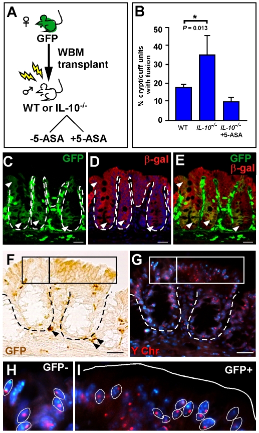
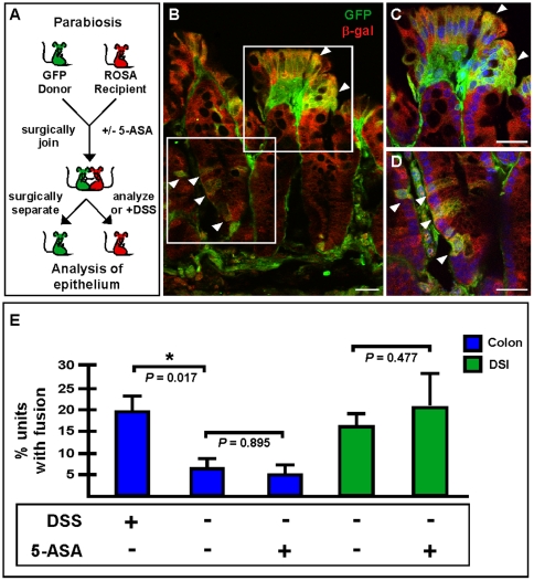
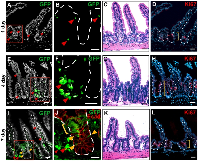
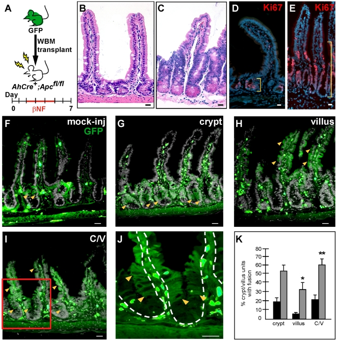
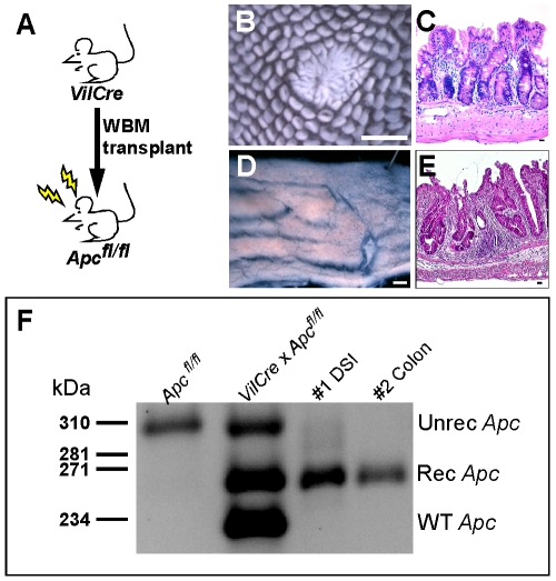
Cell fusion between circulating bone marrow-derived cells (BMDCs) and non-hematopoietic cells is well documented in various tissues and has recently been suggested to occur in response to injury. Here we illustrate that inflammation within the intestine enhanced the level of BMDC fusion with intestinal progenitors. To identify important microenvironmental factors mediating intestinal epithelial cell fusion, we performed bone marrow transplantation into mouse models of inflammation and stimulated epithelial proliferation. Interestingly, in a non-injury model or in instances where inflammation was suppressed, an appreciable baseline level of fusion persisted. This suggests that additional mediators of cell fusion exist. A rigorous temporal analysis of early post-transplantation cellular dynamics revealed that GFP-expressing donor cells first trafficked to the intestine coincident with a striking increase in epithelial proliferation, advocating for a required fusogenic state of the host partner. Directly supporting this hypothesis, induction of augmented epithelial proliferation resulted in a significant increase in intestinal cell fusion. Here we report that intestinal inflammation and epithelial proliferation act together to promote cell fusion. While the physiologic impact of cell fusion is not yet known, the increased incidence in an inflammatory and proliferative microenvironment suggests a potential role for cell fusion in mediating the progression of intestinal inflammatory diseases and cancer.


Conflict of interest statement

Competing Interests: The authors declare commercial funding from Proctor & Gamble Pharmaceuticals and confirm the receipt of this funding does not alter their adherence to all PLoS ONE policies regarding sharing data and materials.

Figures

Figure 1
Figure 1. Inflammation promotes cell fusion between bone marrow-derived cells (BMDCs) and intestinal epithelium.
(A) Schematic representation of experimental design. Whole bone marrow (WBM) from a female GFP-expressing donor mouse was transplanted into lethally irradiated wild-type (WT) or IL-10−/− male mice. A subset of IL-10−/− recipient mice were given the anti-inflammatory drug, 5-ASA. (B) Comparison of cell fusion in colonic epithelium between recipient mice. Cell fusion is quantified as the percentage of crypt/cuff units with at least one GFP-expressing cell. (C–E) Single plane confocal image of a colon cross-section from a ROSA mouse transplanted with GFP WBM. GFP expression (C, green) and β-gal expression (D, red) exist in the same cell (E, yellow) indicating fusion between the donor and recipient cell. Arrowheads denote fused epithelium on the cuff and in the crypts. (F–I) GFP-expressing epithelial cells in transplanted IL-10−/− colons are also fusion products, as determined by co-expression of GFP (F, brown, right box) and the Y chromosome (G, red, right box). (H & I) Higher magnification of GFP-negative and GFP-positive boxed regions from panels F & G. Y chromosome is found in Hoechst stained nuclei (blue, examples circled in white). Solid white line denotes epithelial/luminal border; dashed white lines indicate epithelial/mesenchymal border. Bars = 25 µm.
Figure 2
Figure 2. Intestinal cell fusion persists at low levels in a non-damage model system.
(A) Schematic representation of parabiosis experimental design. GFP and ROSA mice were surgically joined. (B–E) Extensive cell fusion was observed in colons from DSS-treated animals. (B) Single plane confocal microscopy images of GFP (green) and β-galactosidase (red) detected by antibodies demonstrate fusion by co-localization in yellow. Arrowheads denote fused cells. (C & D) depict higher magnifications of the boxed regions in panel B. Nuclei were visualized with the Hoechst dye (blue). Bars = 25 µm. (E) Cell fusion in DSS-treated animals was significantly increased over non-treated animals (P = 0.017). When the animals were given 5-ASA during parabiosis to inhibit inflammation, there was no difference in fusion levels in colon (blue bars; P = 0.895) or DSI (green bars; P = 0.477), however, a baseline level of fusion existed in both tissues.
Figure 3
Figure 3. Increased epithelial proliferation occurred after gamma-irradiation.
Wild-type (WT) mice were transplanted with GFP-expressing whole bone marrow (WBM). The distal small intestine was analyzed at 24 h increments for 1 week. (A–B) At 1 day post-transplantation, few GFP-positive cells (green) were located in the mesenchyme (arrowheads) and none were found in the epithelium. (C–D) H&E and Ki67 detection (red) with Hoechst dye (blue) indicated normal morphology at one day post-transplant. (E–F) At 4 days post-transplant, more GFP-positive cells were found surrounding the crypt (arrowheads), while none were detected in the epithelium. (G–H) H&E and Ki67 staining (red) revealed a dramatic increase in proliferation of the crypts. (I–J) By 7 days post-transplant, single plane confocal microscopy depicts the presence of GFP-positive cells in the mesenchyme surrounding the crypt as well as in the villi core (red arrowheads). GFP-positive epithelium was observed in the stem cell (yellow arrowheads) and transient-amplifying (yellow bracket) zones of crypts. Epithelial cells are marked with antibodies against E-cadherin (red). (K–L) H&E and Ki67 staining depicted morphology close to normal by 7 days post-transplant. Dashed white lines indicate epithelial/mesenchymal border. Red boxes in (A,E,I) are displayed in higher magnification in (B,F,J). Yellow brackets denote the depth of the Ki67-positive cells in (C,G,K). The nuclear dye Hoechst is depicted in grayscale in (A,E,I) and in blues in (D,H,L). D & H are the same tissue sections as A & E, respectively. Bars = 25 µm.
Figure 4
Figure 4. Increased epithelial proliferation correlates with increased cell fusion.
(A) Schematic representation of experimental design. AhCre+;Apcfl/fl mice were transplanted with GFP-expressing whole bone marrow (WBM) on day 0. Two days later, Cre recombinase was induced by β-naphthoflavone (β-NF) administration for 4 consecutive days (days 2–5). Mice were sacrificed on day 7 and the distal small intestine analyzed for fusion (B–E). The intestinal-specific deletion of Apc resulted in an extensive hyperproliferation of crypt cells compared to wild-type (WT) mice, as seen by H&E stain (C vs. B) and Ki67 staining (red, indicated by yellow brackets; E vs. D). (F–K) Detection of GFP-expressing cells (green; yellow arrowheads mark examples) denoting cell fusion was increased in the AhCre+;Apc−/− mice compared to mock-injected WT mice. Three patterns of cell fusion were observed: (G) crypt-only, (H) villus-only, (I) both crypt and villus regions in one crypt/villus unit. Panel (J) is a higher magnification of the red box in panel (I) demonstrating that the Paneth cell region at the base of the crypt remained GFP-negative. Solid white lines denote epithelial/luminal border; dashed white lines indicate epithelial/mesenchymal border. Bars = 25 µm. (K) A significant increase in fusion was observed in villus only (P = 0.011) and crypt/villus (P = 0.009) AhCre+;Apc−/− mice (gray bars) compared to mock-injected WT mice (black bars).
Figure 5
Figure 5. Bone-marrow/epithelial cell fusion causes genetic reprogramming.
(A) Schematic diagram of transplantation scheme. Whole bone marrow (WBM) from mice expressing Cre recombinase driven by the intestinal epithelial-specific Villin promoter (VilCre) was transplanted into recipient mice that were homozygous for floxed Apc. Resulting intestinal phenotypes were observed in transplanted mouse intestine by wholemount analysis as polyps (B) in the distal small intestine (DSI) and as thickened unorganized epithelia (D) in the colon. H&E staining confirmed the phenotypic morphology (C,E). Bars in B & D = 1 mm, bars in C & E = 25 µm. (F) To confirm that the phenotype was the result of recombination at the Apc allele, PCR analysis of epithelium from recipient mice using primers that specifically detect the recombined Apc allele was performed. The 258 bp band was present in the transplanted DSI and colon samples, indicating cell fusion by activation of Cre-recombinase.

References

    1. Alvarez-Dolado M, Pardal R, Garcia-Verdugo JM, Fike JR, Lee HO, et al. Fusion of bone-marrow-derived cells with Purkinje neurons, cardiomyocytes and hepatocytes. Nature. 2003;425:968–973. - PubMed
    1. Camargo FD, Finegold M, Goodell MA. Hematopoietic myelomonocytic cells are the major source of hepatocyte fusion partners. J Clin Invest. 2004;113:1266–1270. - PMC - PubMed
    1. Camargo FD, Green R, Capetanaki Y, Jackson KA, Goodell MA. Single hematopoietic stem cells generate skeletal muscle through myeloid intermediates. Nat Med. 2003;9:1520–1527. - PubMed
    1. Corbel SY, Lee A, Yi L, Duenas J, Brazelton TR, et al. Contribution of hematopoietic stem cells to skeletal muscle. Nat Med. 2003;9:1528–1532. - PubMed
    1. Ferrari G, Cusella-De Angelis G, Coletta M, Paolucci E, Stornaiuolo A, et al. Muscle regeneration by bone marrow-derived myogenic progenitors. Science. 1998;279:1528–1530. - PubMed

Publication types