. 2009 May 15;105(10):102027.
doi: 10.1063/1.3116134. Epub 2009 May 19.

Noninvasive, in vivo imaging of the mouse brain using photoacoustic microscopy

Noninvasive, in vivo imaging of the mouse brain using photoacoustic microscopy

Erich W Stein et al. J Appl Phys. .

Abstract

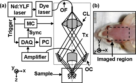
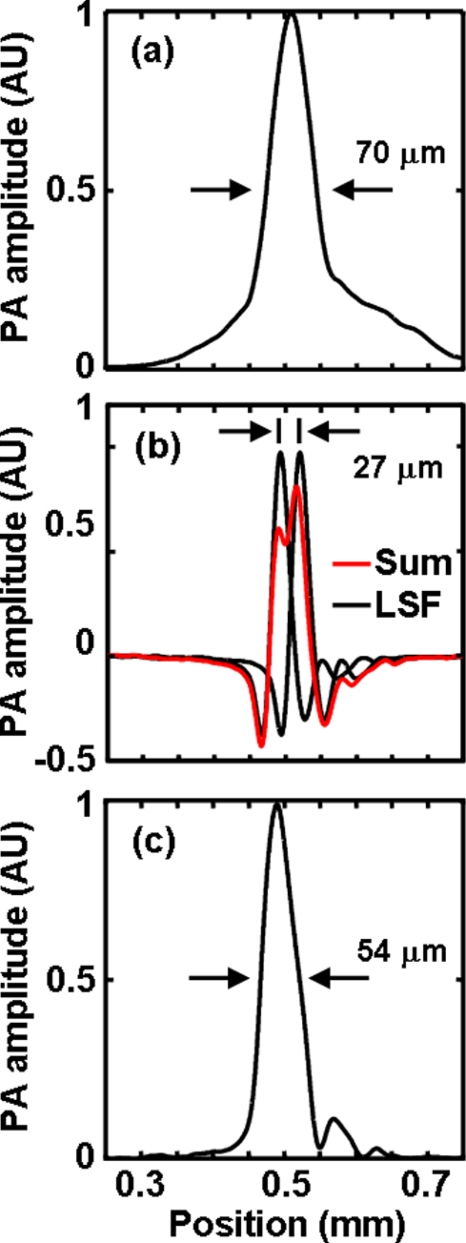
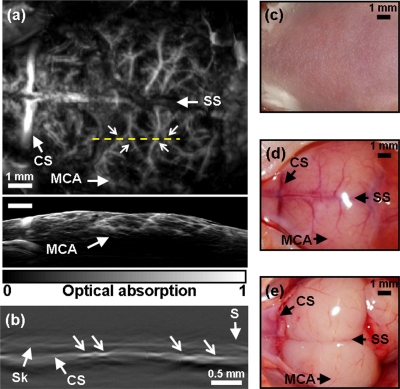
Noninvasive, high resolution imaging of mouse brain activity is poised to provide clinically translatable insights into human neurological disease progression. Toward noninvasive imaging of brain activity through the hemodynamic response, the dark-field photoacoustic microscopy (PAM) technique was enhanced to image the cortex vasculature of the mouse brain in vivo using endogenous hemoglobin contrast. Specifically, the PAM system was redesigned to efficiently collect photoacoustic waves originating from cortical vessels, providing high (70 mum lateral and 54 mum axial) resolution images of the mouse brain vasculature with a contrast-to-noise ratio of 25 dB. These findings confirm the efficacy of PAM to noninvasively image vascular structures in the mouse brain and the potential to image mouse brain function by tracking the hemodynamic response.


Figures

Figure 1
Figure 1
(a) System diagram. OF: optical fiber; CL: conical lens; Tx: transducer; OC: optical condenser; MC: motor controller. (b) Digital photograph of a prepared animal taken prior to imaging, depicting the 10×8 mm2 scanned region.
Figure 2
Figure 2
(a) Transmission coefficient of ultrasound through the rat parietal bone vs ultrasonic frequency and incident angle. LW: predominantly longitudinal wave components; SW: converted shear wave components.
Figure 3
Figure 3
Spatial resolution of the 20 MHz PAM system. (a) Lateral LSF. (b) Shift-and-sum of the nonenveloped axial LSF. (c) Enveloped axial LSF. LSFs obtained by imaging a 6-μm-diameter carbon fiber in optically transparent media.
Figure 4
Figure 4
(a) Noninvasive in vivo PAM image of the mouse cortex vasculature (10×8 mm2). SS: sagittal sinus; MCA: middle cerebral artery; CS: coronal suture. (b) B Scan denoting depth profile along dotted shown line in a. Arrow heads depict cortex vessels viewed in cross section. S: skin surface; Sk: skull; CS: brain cortex surface. (c) Photograph taken prior to imaging, when the cortex vessels are invisible to the naked eye. (d) Photograph taken after image acquisition and scalp removal. (e) Photograph taken after image acquisition and skull removal, showing an unobstructed view of the cortex vessels.

Similar articles

Cited by

References

    1. Hafezparast M., Ahmad-Annuar A., Wood N. W., Tabrizi S. J., and Fisher E. M., Lancet Neurol. 1, 215 (2002). - PubMed
    1. Grinvald A., Lieke E., Frostig R. D., Gilbert C. D., and Wiesel T. N., Nature (London) NATUAS10.1038/324361a0 324, 361 (1986). - DOI - PubMed
    1. Hillman E. M. C., J. Biomed. Opt. 12, 051402 (2007). - PMC - PubMed
    1. Xu M. H. and Wang L. V., Rev. Sci. Instrum. 77, 041101 (2006).
    1. Zhang H. F., Maslov K., Stoica G., and Wang L. V., Nat. Biotechnol. NABIF910.1038/nbt1220 24, 848 (2006). - DOI - PubMed

LinkOut - more resources