. 2009 Aug 4:15:1492-508.

Gap junction remodeling associated with cholesterol redistribution during fiber cell maturation in the adult chicken lens

Affiliations

Gap junction remodeling associated with cholesterol redistribution during fiber cell maturation in the adult chicken lens

Sondip K Biswas et al. Mol Vis. .

Abstract

Purpose: To investigate the structural remodeling in gap junctions associated with cholesterol redistribution during fiber cell maturation in the adult chicken lens. We also evaluated the hypothesis that the cleavage of the COOH-terminus of chick Cx50 (formerly Cx45.6) during fiber cell maturation would affect the gap junction remodeling.

Methods: Freeze-fracture TEM and filipin cytochemistry were applied to visualize structural remodeling of gap junction connexons associated with cholesterol redistribution during fiber cell maturation in adult leghorn chickens (42-62 weeks old). Freeze-fracture immunogold labeling (FRIL) was used to label the specific Cx50 COOH-terminus antibody in various structural configurations of gap junctions.

Results: Cortical fiber cells of the adult lenses contained three subtypes of cholesterol-containing gap junctions (i.e., cholesterol-rich, cholesterol-intermediate, and cholesterol-poor or -free) in both outer and inner cortical zones. Quantitative studies showed that approximately 81% of gap junctions in the outer cortex were cholesterol-rich gap junctions whereas approximately 78% of gap junctions in the inner cortex were cholesterol-free ones. Interestingly, all cholesterol-rich gap junctions in the outer cortex displayed loosely-packed connexons whereas cholesterol-free gap junctions in the deep zone exhibited tightly, hexagonal crystalline-arranged connexons. Also, while the percentage of membrane area specialized as gap junctions in the outer cortex was measured approximately 5 times higher than that of the inner cortex, the connexon density of the crystalline-packed gap junctions in the inner cortex was about 2 times higher than that of the loosely-packed ones in the outer cortex. Furthermore, FRIL demonstrated that while the Cx50 COOH-terminus antibody was labeled in all loosely-packed gap junctions examined in the outer cortex, little to no immunogold labeling was seen in the crystalline-packed connexons in the inner cortex.

Conclusions: Gap junctions undergo significant structural remodeling during fiber cell maturation in the adult chicken lens. The cholesterol-rich gap junctions with loosely-packed connexons in the young outer cortical fibers are transformed into cholesterol-free ones with crystalline-packed connexons in the mature inner fibers. In addition, the loss of the COOH-terminus of Cx50 seems to contribute equally to the transformation of the loosely-packed connexons to the crystalline-packed connexons during fiber cell maturation. This transformation causes a significant increase in the connexon density in crystalline gap junctions. As a result, it compensates considerably for the large decrease in the percentage of membrane area specialized as gap junctions in the mature inner fibers in the adult chicken lens.


Figures

Figure 1
Figure 1
Overview of an intact freeze-fracture replica on the Girder finder grid with index number for systematic examinations of fiber gap junctions in the adult chicken lens. This representative large intact replica was prepared from a mid-sagittal Vibratome lens section (300 μm thick), which was initially trimmed into approximately 2×2 mm blocks to include the superficial and deep cortices from the anterior central region of the lens. Initial identifications of the lens regions (i.e., superficial versus deep) were made and recorded on the intact replica (~1,000 μm in diameter) using the index number. Detailed examinations of the structures of interest were focused primarily on the outer cortex (0-400 μm) and the inner cortex (400-800 μm from the surface) of the lens replica. The scale bar indicates 100 μm.
Figure 2
Figure 2
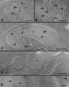
Gap junctions in the outer young cortical fibers (0-400 μm from the surface). A: Both small (arrows) and newly-formed gap junctions (opened arrows) in very superficial cells display loosely-packed connexons. B & C: Several larger gap junction plaques in slightly deeper cells also exhibit loosely-packed connexons. In the images, pf is the P face of the membrane and ef is the E face of the membrane. The scale bars indicate 200 nm.
Figure 3
Figure 3
Gap junctions in the inner mature cortical fibers (400-800 μm from the surface) showing several transitional stages of the structural remodeling of gap junctions. A: In the more superficial regions, small clusters of crystalline-packed connexons (arrows) are seen mixed with other loosely-packed connexons (opened arrows). B: A number of crystalline-packed connexons (arrows) are often aligned into parallel rows within the otherwise loosely-packed (opened arrows) gap junction. C: Another example of a gap junction containing a mixture of crystalline-arranged (arrows) and loosely-packed (opened arrows) connexons in which the crystalline-packed configuration is the major form. In the deeper cortical regions, most connexons are primarily in the tightly- or crystalline-packed arrangements (arrows) in both large (D) and small (E) gap junctions. In the images, pf indicates the P face of membrane and ef indicates the E face of the membrane. The scale bars indicate 200 nm.
Figure 4
Figure 4
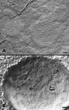
High pressure freezing of gap junctions in cortical fiber cells. Freeze-fracture TEM in conjunction with high pressure freezing shows that while gap junctions display only loosely-packed connexons in superficial cortical fibers (A), a mixture of crystalline-packed (arrows) and loosely-arranged (opened arrow) connexons can be visualized in the deeper cortical fibers of the lens without prior chemical fixation (B). This additional experimental approach is to confirm that the structural remodeling of gap junctions as observed in the chemically fixed lenses in Figure 2 and Figure 3 is a real change during fiber cell maturation. The scale bars indicate 100 nm.
Figure 5
Figure 5
Quantitative analysis of percentage of gap junction population in various size ranges in the outer cortex versus the inner cortex. While the small-size GJs (0.01-0.09 μm2) are mostly populated in both outer and inner cortices, the large-size GJs (0.8-1.75 μm2) are only found in the outer cortical fibers. Importantly, while all small-size GJs in the outer cortex exhibit loosely-packed connexons (Figure 2), the similar small-size GJs in the inner cortex display the crystalline-packed connexons (Figure 3D). This result suggests that (1) small-size newly-formed GJs are gradually developed into large-size ones during fiber cell differentiation in the outer cortex; and (2) large-size GJs undergo structural remodeling (such as removal of cholesterol and re-packing or breakdown of connexons), which results in a significant reduction in the GJ size during maturation and aging in the inner cortex.
Figure 6
Figure 6
Measurements of the percentage of cell membrane area specialized as gap junctions from the outer cortex to the inner cortex. Quantitative measurements of the percentage of cell membrane area specialized as gap junctions from the outer cortex (0-400 μm from surface) to inner cortex (400-800 μm from surface). The area of gap junctions is significantly reduced by approximately 5 times from the outer cortex (~33%) to inner cortex (~6%). A total of 539 junctions were counted in three replicas in the outer cortex, and 141 gap junctions in three replicas in the inner cortex.
Figure 7
Figure 7
Quantitative comparison of the connexon density between the loosely-packed gap junctions in the outer cortex and the crystalline-packed ones in the inner cortex. The number of connexons in the typical crystalline-packed GJs is approximately 2 times higher than that of the loosely-packed GJs. The connexons from fifteen representative gap junctions of each type from three replicas were counted. This result suggests that although the percentage of membrane area specialized as gap junctions is reduced approximately 5 times from the outer cortex to inner cortex (Figure 6), the number of connexons is reduced only about 2.5 times in the deeper cortical region during fiber cell maturation.
Figure 8
Figure 8
Filipin-treated gap junctions. A: A representative cholesterol-rich gap junction with the presence of numerous filipin-cholesterol complexes (FCCs, small arrows) is frequently found in the outer cortical fibers. B and C: Cholesterol-intermediate gap junctions with less number of FCCs (small arrows) are observed in the inner cortical fibers. FCCs (small arrows) are often distributed along patches or parallel rows of crystalline-packed connexons (large arrows). D and E: Cholesterol-free gap junctions are mostly seen in the deeper mature cortical fibers. Note that crystalline-arranged connexons can be visualized more clearly on the E-face (ef) of the membrane. In the images, pf indicates the P-face of the membrane. The scale bars indicate 200 nm.
Figure 9
Figure 9
Freeze-fracture replica immunogold labeling. Freeze-fracture replica immunogold labeling (FRIL) showing the specific labeling of the COOH-terminus of Cx50 (formerly Cx45.6) antibody in many gap junctions in the outer cortical fibers (OC) of the adult chicken lens. The scale bar indicates 500 nm.
Figure 10
Figure 10
Freeze-fracture immunogold labeling (FRIL) of the Cx50 COOH-terminus in different gap junction configurations. FRIL showing the specific labeling of the COOH-terminus of chick Cx50 antibody in gap junctions in the outer cortex (A, B) and in the inner cortex (C-F) at high magnification. In the young outer cortical fibers (OC), immunogold labeling of the Cx50 COOH-terminus antibody can be observed specifically on the P-face (A) and E-face (B) of gap junctions with loosely-packed connexons. However, a considerably smaller number of immunogold labeling (particle) of the Cx50 COOH-terminus antibody is seen in gap junctions with a mixture of crystalline-packed (arrows) and loosely-arranged (open arrows) connexons in the inner cortex (C-F). Note that only a single immunogold particle (arrow) is seen in this GJ with crystalline-packed connexons in (F). For comparison, in the embryonic chick lens (G) in which the gap junctions do not display the distinct crystalline-packed connexons [7], many immunogold particles can be observed in the gap junction with cholesterol-free and non-crystalline-packed connexons. The scale bars indicate 100 nm.
Figure 11
Figure 11
Summary diagram depicting the structural remodeling of gap junctions during fiber cell differentiation and maturation in the adult chicken lens. Stage 1: Formation of small, irregular gap junctions with a few loosely-packed connexons in superficial cortical fiber cells. Stage 2: Growth of a large gap junction with numerous loosely-packed connexons in the young outer cortical fiber cells. Stage 3: Early stage of gap junction remodeling begins in the deeper regions of the outer cortex, displaying several small clusters of crystalline-packed connexons intermingled with a majority of loosely-packed connexons. Stage 4: Mature stage of gap junction remodeling exhibiting distinct hexagonal crystalline-packed connexons observed mainly in the mature inner cortical fiber cells. Due to the compaction of these connexons, these crystalline gap junctions are mostly smaller in size. In concert with these changes, the gap junctions in Stages 1 and 2 are cholesterol-rich, in Stage 3 are cholesterol-intermediate, and in Stage 4 are cholesterol-free. Furthermore, the COOH-terminus of Cx50 is apparently lost in the gap junctions with crystalline-packed connexons in Stage 4.

Similar articles

Cited by

References

    1. Cenedella RJ. Sterol synthesis by the ocular lens of the rat during postnatal development. J Lipid Res. 1982;23:619–26. - PubMed
    1. Zelenka PS. Lens lipids. Curr Eye Res. 1984;3:1337–59. - PubMed
    1. Li LK, So L, Spector A. Membrane cholesterol and phospholipid in consecutive concentric sections of human lenses. J Lipid Res. 1985;26:600–9. - PubMed
    1. Li LK, So L, Spector A. Age-dependent changes in the distribution and concentration of human lens cholesterol and phospholipids. Biochim Biophys Acta. 1987;917:112–20. - PubMed
    1. Li LK, So L. Age dependent lipid and protein changes in individual bovine lenses. Curr Eye Res. 1987;6:599–605. - PubMed

Publication types