

Vascular damage in giant cell arteritis
- PMID: 19657775
- PMCID: PMC4271842
- DOI: 10.1080/08916930903002495
Vascular damage in giant cell arteritis
Abstract
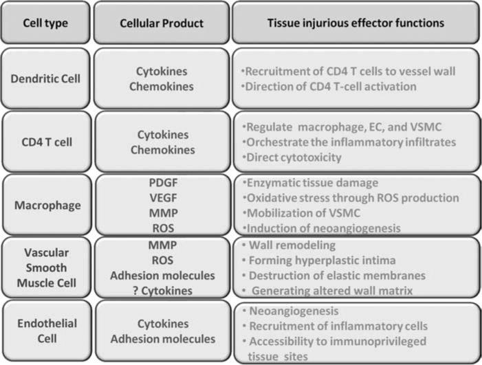
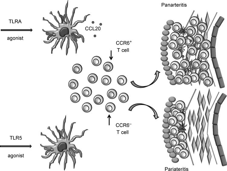
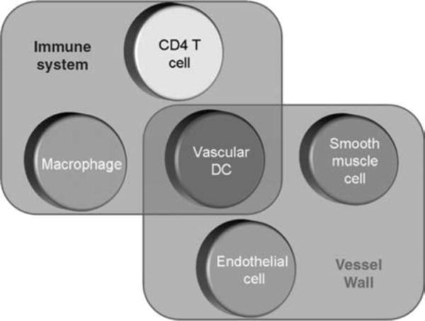
Immune-mediated damage to medium-sized arteries results in wall remodeling with intimal hyperplasia, luminal stenosis and tissue ischemia. In the case of the aorta, vasculitis may result in dissection, aneurysm or rupture. The response-to-injury program of the blood vessel is a concerted action between the immune system and wall-resident cells, involving the release of growth and angiogenic factors from macrophages and giant cells and the migration and hyperproliferation of vascular smooth muscle cells. Innate immune cells, specifically, dendritic cells (DC) positioned in the vessel wall, have been implicated in the earliest steps of vasculitis. Pathogen-derived molecular patterns are capable of activating vascular DC and initiating adaptive immune responses. The pattern of the emerging vessel wall inflammation is ultimately determined by the initial insult. Ligands to toll-like receptor (TLR) 4, such as lipopolysaccharides, facilitate the recruitment of CD4 T cells that invade deep into the wall and distribute in a panarteritic pattern. Conversely, ligands for TLR5 condition vascular DC to support perivasculitic infiltrates. In essence, both innate and adaptive immune reactions collaborate to render the arterial wall susceptible to inflammatory damage. Unique features of the tissue microenvironment, including specialized DC, shape the course of the inflammatory response. Differences in vascular damage pattern encountered in different patients may relate to distinct instigators of vasculitis.
Conflict of interest statement
Figures
References
Publication types
MeSH terms
Substances
Grants and funding
- P30 EY006360/EY/NEI NIH HHS/United States
- R01 AR41974/AR/NIAMS NIH HHS/United States
- R01 AI044142/AI/NIAID NIH HHS/United States
- R01 AR041974/AR/NIAMS NIH HHS/United States
- R01 EY11916/EY/NEI NIH HHS/United States
- P01 HL058000/HL/NHLBI NIH HHS/United States
- R01 HL117913/HL/NHLBI NIH HHS/United States
- R01 AR42527/AR/NIAMS NIH HHS/United States
- R01 AI44142/AI/NIAID NIH HHS/United States
- R01 EY011916/EY/NEI NIH HHS/United States
- U19 AI057266/AI/NIAID NIH HHS/United States
- R01 AR042527/AR/NIAMS NIH HHS/United States
- R01 AG15043/AG/NIA NIH HHS/United States
- U19 AI57266/AI/NIAID NIH HHS/United States
- R01 AG015043/AG/NIA NIH HHS/United States
- P30-EY06360/EY/NEI NIH HHS/United States
LinkOut - more resources
Full Text Sources
Other Literature Sources
Medical
Research Materials