. 2009 Nov;39(11):3000-9.
doi: 10.1002/eji.200838980.

Dual effects of the alloresponse by Th1 and Th2 cells on acute and chronic rejection of allotransplants

Affiliations

Dual effects of the alloresponse by Th1 and Th2 cells on acute and chronic rejection of allotransplants

Ben M Illigens et al. Eur J Immunol. 2009 Nov.

Abstract

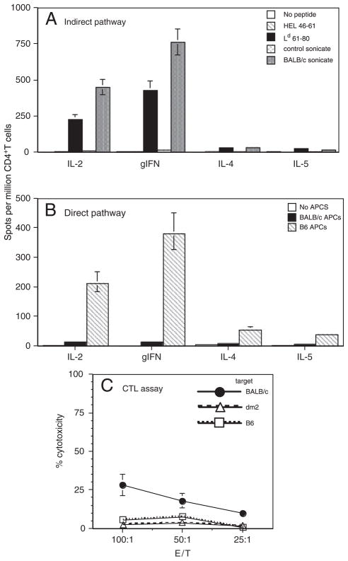
The contribution of direct and indirect alloresponses by CD4(+) Th1 and Th2 cells in acute and chronic rejection of allogeneic transplants remains unclear. In the present study, we addressed this question using a transplant model in a single MHC class I-disparate donor-recipient mouse combination. BALB/c-dm2 (dm2) mutant mice do not express MHC class I L(d) molecules and reject acutely L(d+) skin grafts from BALB/c mice. In contrast, BALB/c hearts placed in dm2 mice are permanently accepted in the absence of chronic allograft vasculopathy. In this model, CD4(+) T cells are activated following recognition of a donor MHC class I determinant, L(d) 61-80, presented by MHC Class II A(d) molecules on donor and recipient APC. Pre-transplantation of recipients with L(d) 61-80 peptide emulsified in complete Freund's adjuvant induced a Th1 response, which accelerated the rejection of skin allografts, but it had no effect on cardiac transplants. In contrast, induction of a Th2 response to the same peptide abrogated the CD8(+) cytotoxic T cells response and markedly delayed the rejection of skin allografts while it induced de novo chronic rejection of heart transplants. This shows that Th2 cells activated via indirect allorecognition can exert dual effects on acute and chronic rejection of allogeneic transplants.


Conflict of interest statement

Conflict of interest: The authors declare no financial or commercial conflict of interest.

Figures

Figure 1
Figure 1
T-cell response of dm2 mice immunized with Ld 61–80 peptide emulsified in CFA. dm2 mice were injected subcutaneously in the hind foootpad with Ld 61–80 peptide (100 ug) emulsified in CFA. Ten days later, draining popliteal LN T cells were collected and tested for cytokine production by ELISPOT and for cytotoxic response against various targets. (A) T cells were cultured in vitro with the immunizing Ag (Ld 61–80, solid bars) or with a control Ag (HEL 46–61, hatched bars). Control non-immunized mice are shown as white bars. The results are presented as spots per million T cells + SD for each cytokine. The results are representative of five mice tested individually. CTL responses of mice immunized with Ld 61–80 peptide (B), no peptide (C), and control OVA 323–339 peptide (D). The ability of T cells to kill allogeneic Ld+ (BALB/c, squares), Ld (B6, circles) and control syngeneic (dm2, triangles) are shown. Spontaneous release ranged from 10 to 15% of maximal release. Data are representative of three independent experiments including 4–5 mice tested individually.
Figure 2
Figure 2
T-cell response of dm2 mice transplanted with a BALB/c skin. dm2 mice (Ld−) were transplanted with a skin from single MHC class I-mismatched BALB/c donors (Ld+). At the time of rejection (10–12 days post-transplantation), CD4+ and CD8+ T cells were isolated from recipients’ spleens and tested for their alloreactivity. (A) CD4+ T cells were tested by ELISPOT for their indirect alloresponse to BALB/c donor. T cells were incubated with recipient APC along with donor sonicates or the dominant donor allopeptide Ld 61–80. T cells cultured with medium alone or control B6 sonicates and irrelevant peptide HEL 46–61 were used as controls. The results are presented as spots per million T cells ± SD for each cytokine. Data are representative of two experiments each including 3–5 mice tested individually. The number of spots obtained with non-transplanted mice ranged from 5–15 spots/million T cells. (B) CD4+ T cells were tested by ELISPOT for their direct alloresponse (MLR) to BALB/c donor. T cells were incubated with donor irradiated MHC class I-mismatched BALB/c APC (solid bars) or fully mismatched B6 APC (hatched bars) or medium (white bars). The results are presented as spots per million T cells ± SD for each cytokine. Data are representative of five experiments each including 2–3 mice tested individually. (C) Cytotoxic response of CD8+ T cells from transplanted dm2 mice against donor BALB/c targets (solid dots). Syngeneic targets (triangles) and third-party B6 targets (squares) were used as controls. The spontaneous release ranged from 10 to 15% of maximal release. The data shown here are representative of two independent experiments including three mice tested individually. The percentages of cytotoxicity recorded with T cells from non-transplanted mice ranged from 2 to 8%.
Figure 3
Figure 3
Schematic representation of the alloresponse in dm2 mice transplanted with BALB/c skin. Donor MHC class I, including Ld 61–80 peptide, are processed and presented in an MHC class II context by recipient APC (exogenous processing, indirect pathway) or donor APC (endogenous processing, direct pathway) to CD4+ T cells. Activated donor-specific CD4+ T cells produce IL-2 thus promoting the differentiation of cytotoxic CD8+ T cells recognizing donor MHC class I at the surface of donor cells (direct allorecognition).
Figure 4
Figure 4
Effects of Ld 61–80 immunization in CFA on allocytotoxicity and skin graft rejection. dm2 mice were immunized subcutaneously with Ld 61–80 MHC class I peptide emulsified in CFA. Ten days later, these mice received a skin graft from a BALB/c donor. (A) Cytotoxic response of CD8+ T cells against donor BALB/c targets (solid circles). Syngeneic targets (triangles) and third-party B6 targets (squares) were used as controls. Spontaneous release ranged from 10 to 15% of maximal release. The percentages of cytotoxicity recorded with T cells from non-transplanted mice ranged from 5 to 10%. (B) Survival of BALB/c skin grafts in Ld 61–80-CFA-immunized dm2 mice (solid line) and control non-immunized mice (dotted line). Data are representative of three independent experiments each including four mice tested individually. The Student’s t-test was used to assess statistical significance between two groups, and one-way ANOVA was used to assess statistical significance between the scores. Graft survival was analyzed using the Kaplan–Meier method, and survival curves were compared using the log-rank test.
Figure 5
Figure 5
Effects of Ld 61–80 immunization in IFA on allocytotoxicity and skin graft rejection. dm2 mice were immunized i.p. with Ld 61–80 MHC class I peptide emulsified in IFA. Ten days later, these mice received a skin graft from a BALB/c donor. (A) Cytotoxic response of CD8+ T cells against donor BALB/c targets (solid circles). Syngeneic targets (triangles) and third-party B6 targets (squares) were used as controls. Spontaneous release ranged from 10 to 15% of maximal release. The percentages of cytotoxicity recorded with T cells from non-transplanted mice ranged from 3 to 7%. (B) Survival of BALB/c skin grafts in Ld 61–80-CFA-immunized dm2 mice (black, solid and dotted lines) and control non-immunized mice (grey doted line). Data are representative of three independent experiments each including three mice tested individually. The Student’s t-test was used to assess statistical significance between two groups, and one-way ANOVA was used to assess statistical significance between the scores. Graft survival was analyzed using the Kaplan–Meier method, and survival curves were compared using the log-rank test.
Figure 6
Figure 6
Histopathology of BALB/c heart transplants from dm2 mice sensitized with Ld 61–80 peptide emulsified in IFA. dm2 mice were immunized i.p. with Ld 61–80 MHC class I peptide emulsified in IFA. Ten days later, these mice received a heart transplant from a BALB/c donor. (A, B) Histology of BALB/c heart transplants from control unimmunized dm2 mice. (C, D) Photomicrographs (40×) of allogeneic hearts harvested 50 and 100days post-transplantation from a Ld 61–80-sensitized recipient mouse display a lymphocytic inflammatory cell infiltrate and vessel obstruction. Data are representative of eight mice tested individually.

References

    1. Krensky AM. T cells in autoimmunity and allograft rejection. Kidney Int Suppl. 1994;44:S50–S56. - PubMed
    1. Lombardi G, Lechler R. The molecular basis of allorecognition of major histocompatibility complex molecules by T lymphocytes. Ann Ist Super Sanita. 1991;27:7–14. - PubMed
    1. Benichou G, Takizawa PA, Olson CA, McMillan M, Sercarz EE. Donor major histocompatibility complex (MHC) peptides are presented by recipient MHC molecules during graft rejection. J Exp Med. 1992;175:305–308. - PMC - PubMed
    1. Fangmann J, Dalchau R, Fabre JW. Rejection of skin allografts by indirect allorecognition of donor class I major histocompatibility complex peptides. Transplant Proc. 1993;25:183–184. - PubMed
    1. Liu Z, Colovai AI, Tugulea S, Reed EF, Fisher PE, Mancini D, Rose EA, et al. Indirect recognition of donor HLA-DR peptides in organ allograft rejection. J Clin Invest. 1996;98:1150–1157. - PMC - PubMed

Publication types

Substances