. 2009 Oct;27(10):2405-13.
doi: 10.1002/stem.191.

Stem-like ovarian cancer cells can serve as tumor vascular progenitors

Affiliations

Stem-like ovarian cancer cells can serve as tumor vascular progenitors

Ayesha B Alvero et al. Stem Cells. 2009 Oct.

Abstract

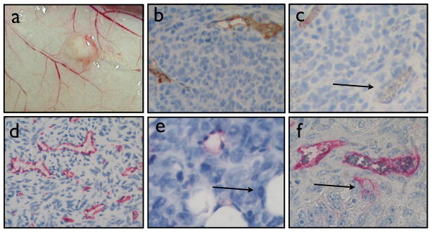
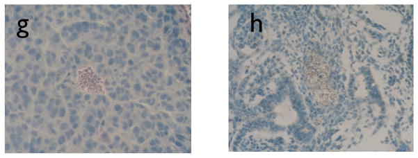
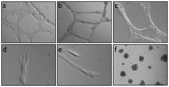
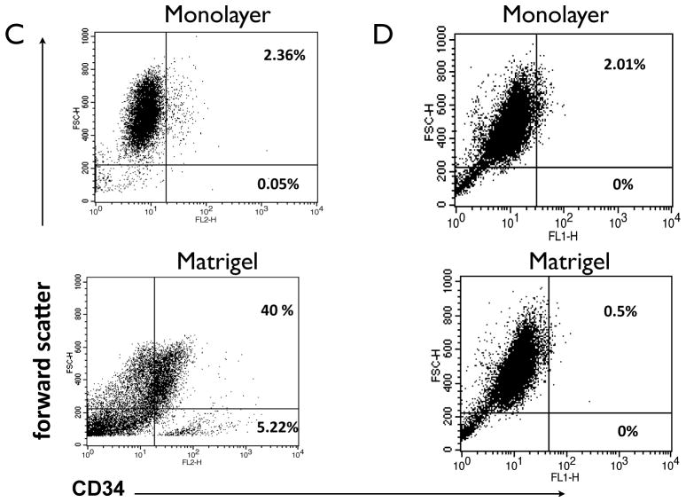
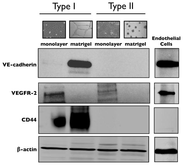
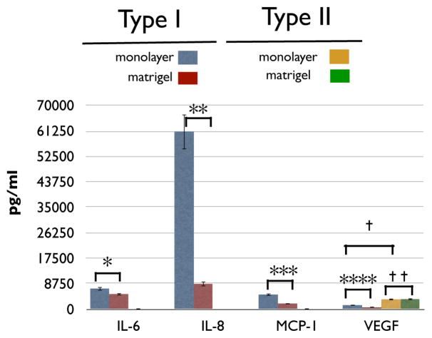
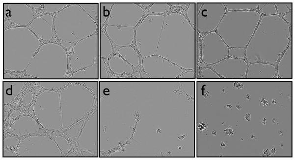
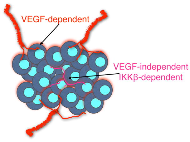
Neovascularization is required for solid tumor maintenance, progression, and metastasis. The most described contribution of cancer cells in tumor neovascularization is the secretion of factors, which attract various cell types to establish a microenvironment that promotes blood vessel formation. The cancer stem cell hypothesis suggests that tumors are composed of cells that may share the differentiation capacity of normal stem cells. Similar to normal stem cells, cancer stem cells (CSCs) have the capacity to acquire different phenotypes. Thus, it is possible that CSCs have a bigger role in the process of tumor neovascularization. In this study, we show the capacity of a specific population of ovarian cancer cells with stem-like properties to give rise to xenograft tumors containing blood vessels, which are lined by human CD34+ cells. In addition, when cultured in high-density Matrigel, these cells mimic the behavior of normal endothelial cells and can form vessel-like structures in 24 hours. Microscopic analysis showed extensive branching and maturation of vessel-like structures in 7 days. Western blot and flow cytometry analysis showed that this process is accompanied by the acquisition of classic endothelial markers, CD34 and VE-cadherin. More importantly, we show that this process is vascular endothelial growth factor-independent, but IKK beta-dependent. Our findings suggest that anti-angiogenic therapies should take into consideration the inherent capacity of these cells to serve as vascular progenitors.


Figures

Figure 1
Figure 1. Xenograft tumors obtained from Type I EOC cells contain both CD34+ (human derived) and CD31+ (mouse derived) blood vessels
(a) Subcutaneous tumors obtained from human ovarian cancer cells with stem-like properties show recruitment of mouse blood vessels. Pure population of Type I EOC cells was injected s.c. in NcR mice. Mice were sacrificed when tumors reached ~8–10mm. Note the projection of host blood vessels towards the human-derived tumor. (b–c) Immunohistochemistry analysis of paraffin sections from xenografts using anti-mouse CD31 antibody. Arrow in C shows CD31-negative blood vessel. (d–f) Immunohistochemistry analysis of paraffin sections from xenografts using anti-human CD34. Arrow in e shows CD34-negative blood vessel; arrow in f shows CD34+ cancer cell. (g) human ovarian cancer tumor stained with mouse anti-CD31. (h) mouse endometrium stained with human anti-CD34. Representative figures of at least 6 independent experiments, n=6 per experiment.
Figure 2
Figure 2. Differentiation of Type I EOC cells into vessel-like structures in vitro. (
a) Human endometrial endothelial cells, (b–e) Type I EOC cells, and (f) Type II EOC cells were plated in high-density Matrigel and vessel formation was monitored for 72h. Note the maturation of branches in d and e. Representative figures of three cell types in each group. Each experiment was repeated at least three times.
Figure 3
Figure 3. Type I EOC cells do not express the endothelial precursor cell markers CD133 and CD34 but acquired CD34 after differentiation
Cultures of Type I EOC cells grown in monolayer were analyzed for the expression of (a) CD133 and (b) CD34 by flow cytometry. Note that the cells are negative for the two markers. (c) CD34 levels were compared in Type I EOC cells grown for 72h either in monolayer or Matrigel. A significant increase on CD34 + cells is observed on differentiated Type I EOC cells. (d) CD34 levels were compared in Type I EOC cells grown for 72h either in monolayer or Matrigel. The CD34 antigenic shift was not observed in Type II EOC cells. Representative experiment done with three different clones.
Figure 4
Figure 4. Type I EOC cells acquire endothelial cell markers following differentiation in Matrigel
Cells were grown for 72h either as a monolayer or in Matrigel and cell lysates analyzed by western blot for the endothelial-specific marker VE-cadherin, the stem cell marker CD44, and VEGFR-2.
Figure 5
Figure 5. Levels of cytokines secreted by Type I or II EOC cells cultures in monolayer or Matrigel
Cultures were grown for 72h either as monolayer or in Matrigel and cell-free supernatants were used to measure the levels of cytokines/chemokines as described in the Materials and Methods. * p = 0.03; ** p = 0.01; *** p = 0.07; **** p = 0.01; † p = 0.005; † p = 0.01.
Figure 6
Figure 6. Type I EOC cell vessel formation is independent on external growth factors but dependent on IKKβ
(a–e) Type I EOC cells in matrigel: control, in growth factor-reduced Matrigel, in 50% Type II conditioned media, with 1 μg/ml sFlt-1, and with 0.4μM BAY 11-7082, respectively; (f–g) Type II EOC cells in matrigel: control, and in 50% Type I conditioned media, respectively; (h–k) normal human endometrial endothelial cells in Matrigel: control, in growth factor-reduced matrigel, with 1 μg/ml sFlt-1, with 0.4μM BAY 11-7082, respectively.
Figure 7
Figure 7. Model showing contribution of Type I EOC cells in tumor neovascularization
We propose that in ovarian cancer, neovascularization can occur through classical VEGF-dependent angiogenesis and more importantly, through a VEGF-independent but IKKβ-dependent differentiation of Type I EOC cells.

Similar articles

Cited by

References

    1. Sato Y. Molecular diagnosis of tumor angiogenesis and anti-angiogenic cancer therapy. Int J Clin Oncol. 2003;8:200–206. - PubMed
    1. Folkman J, Shing Y. Angiogenesis. J Biol Chem. 1992;267:10931–10934. - PubMed
    1. Rafii S. Circulating endothelial precursors: mystery, reality, and promise. J Clin Invest. 2000;105:17–19. - PMC - PubMed
    1. Goh PP, Sze DM, Roufogalis BD. Molecular and cellular regulators of cancer angiogenesis. Curr Cancer Drug Targets. 2007;7:743–758. - PubMed
    1. Cao Y. Tumor angiogenesis and molecular targets for therapy. Front Biosci. 2009;14:3962–3973. - PubMed

Publication types

MeSH terms