. 2009 Dec;22(6):773-84.
doi: 10.1111/j.1755-148X.2009.00616.x. Epub 2009 Jul 30.

NRG1 / ERBB3 signaling in melanocyte development and melanoma: inhibition of differentiation and promotion of proliferation

Affiliations

NRG1 / ERBB3 signaling in melanocyte development and melanoma: inhibition of differentiation and promotion of proliferation

Kristina Buac et al. Pigment Cell Melanoma Res. 2009 Dec.

Abstract

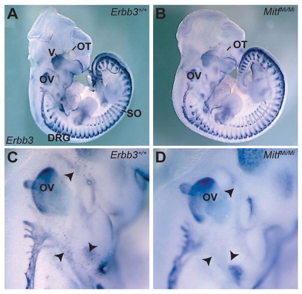
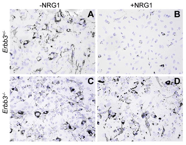
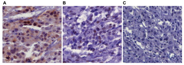
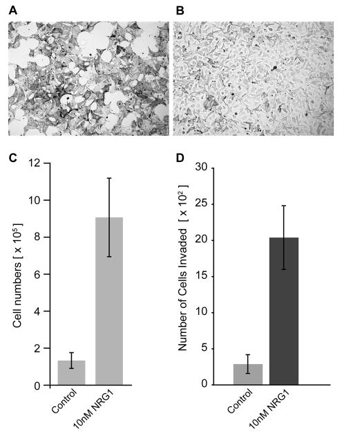
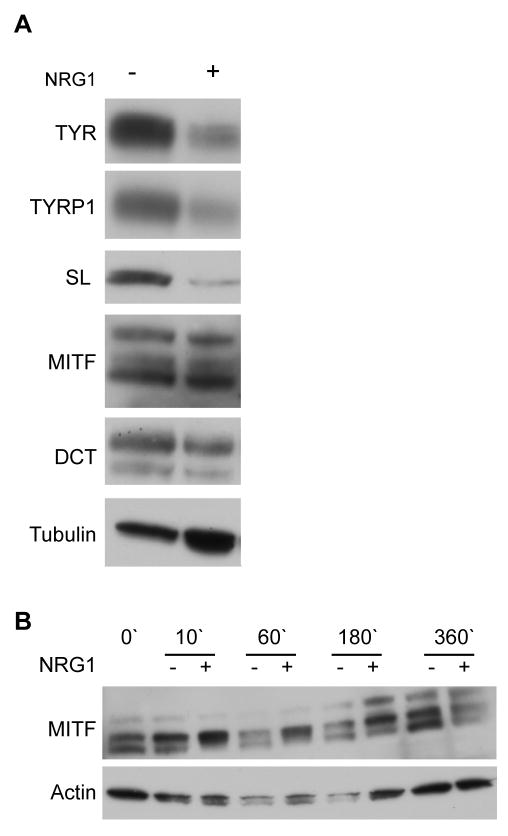
Neuregulin (NRG) signaling through the receptor tyrosine kinase, ERBB3, is required for embryonic development, and dysregulated signaling has been associated with cancer progression. Here, we show that NRG1/ERBB3 signaling inhibits melanocyte (MC) maturation and promotes undifferentiated, migratory and proliferative cellular characteristics. Embryonic analyses demonstrated that initial MC specification and distribution were not dependent on ERBB3 signaling. However NRG1/ERBB3 signaling was both necessary and sufficient to inhibit differentiation of later stages of MC development in culture. Analysis of tissue arrays of human melanoma samples suggests that ERBB3 signaling may also contribute to metastatic progression of melanoma as ERBB3 was phosphorylated in primary tumors compared with nevi or metastatic lesions. Neuregulin 1-treated MCs demonstrated increased proliferation and invasion and altered morphology concomitant with decreased levels of differentiation genes, increased levels of proliferation genes and altered levels of melanoma progression and metastases genes. ERBB3 activation in primary melanomas suggests that NRG1/ERBB3 signaling may contribute to the progression of melanoma from benign nevi to malignancies. We propose that targeting ERBB3 activation and downstream genes identified in this study may provide novel therapeutic interventions for malignant melanoma.


Figures

Figure 1
Figure 1. Erbb3is expressed in MC precursors during embryonic development
Lateral view of Erbb3 whole-mount in situ hybridization in wild-type (A, C) and MitfMi/Mi (B, D) embryos at E11.5. In both wild-type embryos and MitfMi/Mi mutants, Erbb3 expression is observed in the cranial ganglia and nerves, DRG and somites. In wild-type embryos (C), a population of Erbb3-expressing cells (black arrowheads) is detected in the vicinity of the otic vesicle, the location where Mbs are normally found. These Erbb3-expressing cells were absent in Mb-deficient mutants (D), indicating that these cells are Mbs. Labeled structures are otocyst (OT) trigeminal ganglia (V), dorsal root ganglia (DRG), otic vesicle (OV) and somites (SO).
Figure 2
Figure 2. MC differentiation in NC cultures
Bright field images of Erbb3+/- (A,B) and Erbb3-/- (C,D) NC cultures grown in the absence (A,C) or presence (B,D) of NRG1. Cells were cultured for two weeks after neural tube isolation, replated and DAPI stained one day later. Although differentiated MCs (dark cells) were detected in both genotypes, pigmented MCs from Erbb3 mutant cultures (C) were darker. When exogenous NRG1 was added to Erbb3+/- NC cultures (B), a dramatic reduction in pigmented MCs was observed. No such difference was noted when Erbb3 mutant cultures (D) were grown with NRG1, suggesting that activation of NRG1/ERBB3 signaling inhibits MC differentiation.
Figure 3
Figure 3. Phospho-ERBB3 staining in melanoma tissue arrays
Representative photomicrographs of immunohistochemistry for phospho-ERBB3. All images taken at 320× magnification. (A) Nuclear and cytoplasmic staining of phospho-ERBB3 in a primary melanoma. (B) Nuclear staining of phospho-ERBB3 in a melanoma metastatic to a lymph node. (C) A metastatic melanoma that is negative for staining of phospho-ERBB3.
Figure 4
Figure 4. NRG1 increases proliferation and invasion of melan-Ink4a cells
Bright filed images of melan-Ink4a cells (A, B) in media lacking TPA and CT (A) and in media lacking TPA and CT but with 10nM NRG1 added (B). (C) Cell doublings in the absence of TPA and CT (control) and in media containing NRG1 without TPA and CT (10 nM NRG1). Cells were plated at equal numbers (2.5 × 10⁵) and total numbers of cells were counted four days after the indicated treatments using hemocytometer. While melan-Ink4a grown in the absence of TPA/CT stopped proliferating, addition of NRG1 increased number of Melan-Ink4a cells. D) Increased invasion in the presence of NRG1. Control and NRG1-treated Melan-Ink4a cells were grown in Boyden chambers and assessed for their ability to invade through basement membrane matrix. For quantification, stained cells were photographed on Zeiss Axiovert 135 microscope and cells were counted in at least 16 random fields. Columns represent the absolute number of cells that invaded the matrix 48 hours after seeding (n=4).
Figure 5
Figure 5. NRG1 effects expression of melanogenic enzymes
(A) Protein levels of Tyrosinase (TYR), Tyrosinase-related protein 1 (TYRP1) and Silver (SL) were downregulated in NRG1-treated cultures (22 days after treatment). MITF and DCT expression was not visibly altered by NRG1. (B) A time-course analysis of MITF expression at 10, 60, 180, and 360 minutes after addition of NRG1. MITF phosphorylation (upper MITF band) was increased by NRG1 at 180′ and protein levels were decreased by NRG1 after 360 minutes, indicating that activation of ERBB3 pathway can regulate MITF activity levels.

References

    1. Baxter LL, Hsu BJ, Umayam L, Wolfsberg TG, Larson DM, Frith MC, Kawai J, Hayashizaki Y, Carninci P, Pavan WJ. Informatic and genomic analysis of melanocyte cDNA libraries as a resource for the study of melanocyte development and function. Pigment Cell Res. 2007;20:201–9. - PubMed
    1. Baxter LL, Pavan WJ. Pmel17 expression is Mitf-dependent and reveals cranial melanoblast migration during murine development. Gene Expr Patterns. 2003;3:703–7. - PubMed
    1. Bennett DC. Colour genes, oncogenes and melanocyte differentiation. J Cell Sci. 1991;98(Pt 2):135–9. - PubMed
    1. Bennett DC. How to make a melanoma: what do we know of the primary clonal events? Pigment Cell Melanoma Res. 2008;21:27–38. - PubMed
    1. Bennett DC, Trayner ID, Piao X, Easty DJ, Kluppel M, Alexander WS, Wagner EF, Bernstein A. recessive spotting: a linked locus that interacts with W/Kit but is not allelic. Genes Cells. 1998;3:235–44. - PubMed

Publication types

MeSH terms