. 2009 Jul;157(1):71-82.
doi: 10.1111/j.1365-2249.2009.03945.x.

Up-regulation of CC chemokine receptor 6 on tonsillar T cells and its induction by in vitro stimulation with alpha-streptococci in patients with pustulosis palmaris et plantaris

Affiliations

Up-regulation of CC chemokine receptor 6 on tonsillar T cells and its induction by in vitro stimulation with alpha-streptococci in patients with pustulosis palmaris et plantaris

T Yoshizaki et al. Clin Exp Immunol. 2009 Jul.

Abstract

Pustulosis palmaris et plantaris (PPP) is a tonsil-related disease; tonsillectomy is somewhat effective in treating the condition. However, the aetiological association between the tonsils and PPP has not yet been elucidated fully. Recently, some chemokines and chemokine receptors, including CC chemokine receptor (CCR) 4, CCR6 and CX chemokine receptor (CXCR) 3, have been reported to play important roles in the development of psoriasis, a disease related closely to PPP. In this study, we found that CCR6 expression on both tonsillar and peripheral blood T cells was up-regulated more intensively in PPP patients than in non-PPP patients (P < 0.001 for both), but CCR4 and CXCR3 expressions were not. In vitro stimulation with alpha-streptococcal antigen enhanced CCR6 expression significantly on tonsillar T cells in PPP patients (P < 0.05), but this was not observed in non-PPP patients. The chemotactic response of tonsillar T cells to the CCR6 ligand CC chemokine ligand (CCL) 20 was significantly higher in PPP patients than in non-PPP patients (P < 0.05). The percentage of CCR6-positive peripheral blood T cells decreased after tonsillectomy in PPP patients (P < 0.01); this decrease correlated with an improvement of skin lesions (P < 0.05, r = -0.63). The numbers of CCR6-positive cells and the expression of CCL20 were increased significantly in pathological lesions compared with non-pathological lesions in PPP skin (P < 0.01, P < 0.05 respectively). These results suggest that a novel immune response to alpha-streptococci may enhance CCR6 expression on T cells in tonsils and that CCR6-positive T cells may move to peripheral blood circulation, resulting in recruitment to target skin lesions expressing CCL20 in PPP patients. This may be one of the key roles in pathogenesis of the tonsil-related disease PPP.


Figures

Fig. 1
Fig. 1
Representative findings of palmar skin in patients with pustulosis palmaris et plantaris (PPP) before (a) and 6 months after tonsillectomy (b) in patient no. 9. The skin severity score was 1 point, and the efficacy of the tonsillectomy was judged to be ‘markedly effective’.
Fig. 2
Fig. 2
Percentages of CC chemokine receptor (CCR) 4/CD3 (a), CCR6/CD3 (b) and CXCR3/CD3 (c) double-positive cells among tonsillar CD3-positive cells from PPP patients and non-PPP patients. Tonsillar mononuclear cells (TMCs) were isolated from PPP patients (n = 14) and non-PPP patients (n = 12) with recurrent tonsillitis (RT) by tonsillectomy. Two-colour flow cytometric analysis revealed that the percentage of CCR6/CD3 double-positive cells among CD3-positive cells from TMCs was significantly higher in PPP patients than in non-PPP patients (P < 0·001). The median values are displayed as short bars (–). The Mann–Whitney U-test was used to determine the P-values.
Fig. 3
Fig. 3
Percentages of CC chemokine receptor (CCR) 4/CD3 (a), CCR6/CD3 (b) and CXCR3/CD3 (c) double-positive cells among peripheral blood CD3-positive cells from PPP patients and non-PPP patients. Peripheral blood mononuclear cells (PBMCs) were isolated from PPP patients before tonsillectomy (n = 14) and from normal volunteers (NV) (n = 12). Two-colour flow cytometric analysis revealed that the percentage of CCR6/CD3 double-positive cells among CD3-positive cells from PBMCs was significantly higher in PPP patients than in non-PPP patients (P < 0·001). The median values are displayed as short bars (–). The Mann–Whitney U-test was used to determine the P-values.
Fig. 4
Fig. 4
Changes in percentage of CC chemokine receptor (CCR) 6/CD3 double-positive cells among tonsillar CD3-positive cells by in vitroα-streptococcal stimulus (a) and changes in CCR6 stimulation rate (b). (a) Tonsillar mononuclear cells (TMCs) from PPP patients (n = 7) and non-PPP patients with recurrent tonsillitis (RT) (n = 7) were cultured for 3 days in the absence or presence of phytohaemagglutinin (PHA) or α-streptococcal antigens. Each value is expressed as a percentage of CD3/CCR6 double-positive cells among CD3-positive cells and analysed by two-color flow cytometry. PHA stimulation increased the percentage of CCR6/CD3 double-positive cells among CD3-positive cells in both PPP and non-PPP patients (each P < 0·05). Stimulation with Streptococcus mitis antigen increased the percentage of CCR6/CD3 double-positive cells among CD3-positive cells in PPP patients (P < 0·05) but not in non-PPP patients. Following stimulation with S. salivarius antigen, the percentage of CCR6/CD3 double-positive cells among CD3-positive cells was significantly higher in PPP patients than in non-PPP patients (P < 0·05). (b) The CCR6 stimulation rate was calculated in the following manner: (the percentage of CCR6/CD3 double-positive cells among CD3-positive cells with stimulus/that without stimulus) × 100. The CCR6 stimulation rate with PHA stimulus was not different between PPP (n = 7) and non-PPP patients (n = 7). However, the CCR6 stimulation rates observed with S. mitis and S. salivarius were significantly higher in PPP patients than in non-PPP patients (P < 0·05 for both). The median values are displayed as short bars (–). The Mann–Whitney U-test was used to determine the P-values.
Fig. 5
Fig. 5
Chemotactic response of tonsillar T cells to CCC chemokine ligand (CCL) 20. CD3-positive cells were isolated from TMCs of both PPP (n = 7) and non-PPP patients (n = 7) using a magnetic bead method. One million CD3-positive cells were added to the top chamber with a 3-µm pore size polycarbonate transwell culture insert and incubated at three levels of concentration with recombinant CCL20 in the bottom chamber for 3 h at 37°C. The number of cells in the bottom chamber was counted. The chemotaxis index was calculated as the ratio of the number of cells migrating to the bottom chamber divided by the number of migrating cells in the bottom chamber without CCL20. The chemotaxis index was significantly higher in PPP patients than in non-PPP patients at the concentration of 100 ng/ml CCL20 (P < 0·01). The median values are displayed as short bars (–). The Mann–Whitney U-test was used to determine the P-values.
Fig. 6
Fig. 6
Changes in percentage of CC chemokine receptor (CCR) 6/CD3 double-positive cells among peripheral blood CD3-positive cells in PPP patients after tonsillectomy. (a) The percentage of CCR6/CD3 double-positive cells among CD3-positive cells decreased significantly at 6 months after tonsillectomy (n = 12, P < 0·01). (b) The rate of decrease of CCR6 expression rate was estimated as follows: [(the percentage of CCR6/CD3 double-positive cells among CD3-positive cells before tonsillectomy) – (that after tonsillectomy)]/(that before tonsillectomy) × 100%. There was a significant correlation between the skin severity score after tonsillectomy and the rate of decrease of CCR6 (P < 0·05, r = −0·63). The median values are displayed as short bars (–). The Wilcoxon signed rank test and the Spearman's rank correlation coefficient were used to determine the P-values.
Fig. 7
Fig. 7
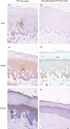
Representative immunohistological profiles of CD3 (a, b), CC chemokine receptor (CCR) 6 (c, d) and CC chemokine ligand (CCL) 20 (e, f) staining in pathological (a, c, d) and non-pathological lesions (b, d, e) from plantar biopsy specimens of PPP patients (×200). Numerous CD3- (a) and CCR6-positive cells (c) were found around pustules in the epidermis and papillary dermis in pathological lesions. However, in non-pathological lesions, CD3- and CCR6-positive cells were rarely found (b, d). CCL20-positive cells with strong immunoreactivity were found in the epidermis (e), but very few CCL20-positive cells were found in the epidermis (f).

References

    1. Andrews GC, Machacek GF. Recalcitrant pustular eruptions of the palms and soles. Arch Dermatol. 1934;29:548–62.
    1. Hellgren L, Mobacken H. Pustulosis palmaris et plantaris. Prevalence, clinical observations and prognosis. Acta Derm Venereol. 1971;51:284–8. - PubMed
    1. Kataura A, Tsubota H. Clinical analyses of focus tonsil and related diseases in Japan. Acta Otolaryngol. 1996;(Suppl 523):161–4. - PubMed
    1. Ono T. Evaluation of tonsillectomy as a treatment for pustulosis palmaris et plantaris. J Dermatol. 1977;4:163–72. - PubMed
    1. Yamanaka N, Shido F, Kataura A. Tonsillectomy-induced changes in anti-keratin antibodies in patients with pustulosis palmaris et plantaris: a clinical correlation. Arch Otorhinolaryngol. 1989;246:109–12. - PubMed

MeSH terms