Review
. 2009 Aug;90(4):367-86.
doi: 10.1111/j.1365-2613.2009.00656.x.

Experimental mouse models for hepatocellular carcinoma research

Affiliations
Review

Experimental mouse models for hepatocellular carcinoma research

Femke Heindryckx et al. Int J Exp Pathol. 2009 Aug.

Abstract

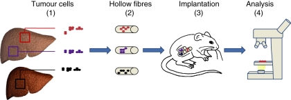
Every year almost 500,000 new patients are diagnosed with hepatocellular carcinoma (HCC), a primary malignancy of the liver that is associated with a poor prognosis. Numerous experimental models have been developed to define the pathogenesis of HCC and to test novel drug candidates. This review analyses several mouse models useful for HCC research and points out their advantages and weaknesses. Chemically induced HCC mice models mimic the injury-fibrosis-malignancy cycle by administration of a genotoxic compound alone or, if necessary, followed by a promoting agent. Xenograft models develop HCC by implanting hepatoma cell lines in mice, either ectopically or orthotopically; these models are suitable for drug screening, although extrapolation should be considered with caution as multiple cell lines must always be used. The hollow fibre assay offers a solution for limiting the number of test animals in xenograft research because of the ability for implanting multiple cell lines in one mouse. There is also a broad range of genetically modified mice engineered to investigate the pathophysiology of HCC. Transgenic mice expressing viral genes, oncogenes and/or growth factors allow the identification of pathways involved in hepatocarcinogenesis.


Figures

Figure 1
Figure 1
Tumour cells derived from human (or mouse) hepatic tumours (1) are placed in small semi-permeable tubes known as hollow fibres (2). The fibres are cultured for 24-48 hours in vitro before subcutaneous or intraperitoneal implantation in nude mice. The cells can be retrieved after the in vivo assay and used for subsequent analysis (4).
Figure 2
Figure 2
c-myc is one of the key elements in the malignang transformation. By (1) preventing differentiation and/or (2) cell cycle arrest, deregulated Myc forces cells to remain in a (3) proliferative state and causes (4) genomic instability. (5) Apoptosis is induced by activating death receptor pathways, in which myc has several points of regulation. c-Myc over-expression results in elevated expression of genes involved in regulating the (6) cellular metabolism, allowing tumour cells to switch to an anaerobic metabolism when oxygen is depleted).
Figure 3
Figure 3
Beta-catenin is an integral componant of the Wnt-signaling pathway, a pathway that is one of the central players in maintaining liver health. The association between beta-catenin and E-cadherin is found in the hepatocyte membrane and has significant implications in (1) cell-cell adhesion. Phosphorylation of beta-catenin inhibits the beta-catenin-E-cadherin association, leading to disruption of adherens junctions and loss of intracellular adhesion. The dissociation of this complex induces nuclear translocation of beta-catenin, leading to target gene expression. Nuclear translocation of beta-catenin is associated with hepatocyte proliferation during development and after partial hepatectomy. Beta-catenin regulates the expression of genes involved in (3) hepatocyte maturation and in (4) biliary specification. The interaction between the protein adenomatous polyposis coli and beta-catenin, and the involvement of beta-catenin in the regulation of proteins important in ammonia metabolism, play an important role in the (5) zonation of the liver, dividing the liver in several structural and (6) metabolic regions.
Figure 4
Figure 4
Transforming growth factor alpha (TGF-alpha) and epidermal growth factor (EGF) are important hepatic mitogens that both bind on the epidermal growth factor receptor (EGFR). Upon activation, EGFR undergoes dimerisation which stimulates its intrinsic intracellular protein-tyrosine kinase activity. This leads to the initiation of several signal transduction cascades, leading to cell growth, proliferation and differentiation.
Figure 5
Figure 5
The fibroblas growth factor 19 (FGF19) is a high affinity ligand for the fibroblas growth factor receptor 4 (FGFR4) expressed in hepatocytes. Binding of FGF19 to FGFR4 leads to an increased cellular metabolism, associated with the production of reactive oxygen species (ROS). Wether HCC formation is an indirect effect of altered metabolism or a direct affect of FGF19 is unknown.
Figure 6
Figure 6
Phosphatase and tensin homolog (PTEN) is a tumour suppressor gene involved in the regulation of the threonine kinase protein kinase B (PKB/akt) pathway, by dephosphorylating phosphatidylinositol (3,4,5)-triphosphate (PIP3). The PKB/akt pathway is involved in the regulation of the cell cycle, apoptosis and cell growth.

References

    1. Araki K, Miyazaki J, Hino O, et al. Expression and replication of hepatitis B virus genome in transgenic mice. Proc. Natl Acad. Sci. USA. 1989;86:207–211. - PMC - PubMed
    1. Araki K, Hino O, Miyazaki J, Yamamura K. Development of 2 types of hepatocellular-carcinoma in transgenic mice carrying the Sv40 large T-antigen gene. Carcinogenesis. 1991;12:2059–2062. - PubMed
    1. Autrup H, Wakhisi J. Detection of exposure to aflatoxin in an African population. IARC Sci. Publ. 1988;8:63–66. - PubMed
    1. Avantaggiati ML, Natoli G, Balsano C, et al. The hepatitis B virus (HBV) pX transactivates the c-fos promoter through multiple cis-acting elements. Oncogene. 1993;8:1567–1574. - PubMed
    1. Avasarala S, Yang L, Sun Y, et al. A temporal study on the histopathological, biochemical and molecular responses of CCl(4)-induced hepatotoxicity in Cyp2e1-null mice. Toxicology. 2006;228:310–322. - PubMed