

Diabetes induces stromal remodelling and increase in chondroitin sulphate proteoglycans of the rat ventral prostate
- PMID: 19659898
- PMCID: PMC2741150
- DOI: 10.1111/j.1365-2613.2009.00657.x
Diabetes induces stromal remodelling and increase in chondroitin sulphate proteoglycans of the rat ventral prostate
Abstract
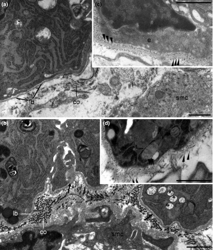
Extracellular matrix (ECM) remodelling is an important process involved in prostate cancer progression. Alterations in ECM caused by diabetes in different tissues such as kidney is well described; however, it is poorly investigated in prostate. The aim of this study was to evaluate changes in ECM of rat prostate showing gland atrophy caused by diabetes and their implications in development of malignant lesions. Diabetes was induced in Wistar rats using alloxan (45 mg/kg bw). After 90 days of diabetes onset, animals were killed and ventral prostate was removed and prepared for light microscopy following immunoreaction for fibronectin, chondroitin sulphate and Picrossirius staining for collagen fibres. Proteoglycans (PG) were identified at transmission electron microscopy after fixation with Cuprolinic Blue. Diabetes led to a thickening of 25% in the acinar basement membrane accompanied by increase and disorganization of its proteoglycans (P1). Three additional populations of prostatic stromal PGs were identified: collagen fibril linked (P2) and interstitial (P3) and (P4) PGs. Diabetes increased P3 and mainly P4 which had higher dimension and accumulated around the smooth muscle cells. In addition, an increase in chondrotin sulphate (33%, mainly in sites where P4 were noted) and collagen (44%) was noted in diabetic rats, whereas fibronectin did not change. Atrophic changes observed in rat ventral prostate after diabetes are accompanied by stromal remodelation related to increase in collagen and chondroitin sulphate proteoglycans. Thus, diabetes can promote a stromal microenvironment rich in elements that could favour cell migration, proliferation and pathological process.
Figures


Similar articles
-
Influence of testosterone on chondroitin sulphate proteoglycan in the rat prostate.Biochem Cell Biol. 1996;74(5):645-51. doi: 10.1139/o96-069. Biochem Cell Biol. 1996. PMID: 9018371
-
Short-term stromal alterations in the rat ventral prostate following alloxan-induced diabetes and the influence of insulin replacement.Micron. 2012 Feb;43(2-3):326-33. doi: 10.1016/j.micron.2011.09.009. Epub 2011 Sep 29. Micron. 2012. PMID: 22014851
-
Functional involvement of sciatic nerve-derived versican- and decorin-like molecules and other chondroitin sulphate proteoglycans in ECM-mediated cell adhesion and neurite outgrowth.Eur J Neurosci. 1995 Apr 1;7(4):805-14. doi: 10.1111/j.1460-9568.1995.tb00683.x. Eur J Neurosci. 1995. PMID: 7620627
-
Mapping of proteoglycans in atherosclerotic lesions.Eur Heart J. 1990 Aug;11 Suppl E:29-40. doi: 10.1093/eurheartj/11.suppl_e.29. Eur Heart J. 1990. PMID: 2226532 Review.
-
Structural variability of large and small chondroitin sulphate/dermatan sulphate proteoglycans.Biochem Soc Trans. 1990 Apr;18(2):209-12. doi: 10.1042/bst0180209. Biochem Soc Trans. 1990. PMID: 2199261 Review. No abstract available.
Cited by
-
Prostate hyperplasia caused by long-term obesity is characterized by high deposition of extracellular matrix and increased content of MMP-9 and VEGF.Int J Exp Pathol. 2015 Feb;96(1):21-30. doi: 10.1111/iep.12107. Epub 2014 Dec 21. Int J Exp Pathol. 2015. PMID: 25529509 Free PMC article.
-
Effect of Melatonin Intake on Oxidative Stress Biomarkers in Male Reproductive Organs of Rats under Experimental Diabetes.Oxid Med Cell Longev. 2015;2015:614579. doi: 10.1155/2015/614579. Epub 2015 May 6. Oxid Med Cell Longev. 2015. PMID: 26064423 Free PMC article.
-
Proliferation and apoptotic rates and increased frequency of p63-positive cells in the prostate acinar epithelium of alloxan-induced diabetic rats.Int J Exp Pathol. 2010 Apr;91(2):144-54. doi: 10.1111/j.1365-2613.2009.00696.x. Epub 2009 Dec 22. Int J Exp Pathol. 2010. PMID: 20041964 Free PMC article.
-
Thiazolidinediones and metformin associated with improved survival of diabetic prostate cancer patients.Ann Oncol. 2011 Dec;22(12):2640-2645. doi: 10.1093/annonc/mdr020. Epub 2011 Mar 17. Ann Oncol. 2011. PMID: 21415239 Free PMC article.
-
Influence of Melatonin on the Proliferative and Apoptotic Responses of the Prostate under Normal and Hyperglycemic Conditions.J Diabetes Res. 2015;2015:538529. doi: 10.1155/2015/538529. Epub 2015 Jul 30. J Diabetes Res. 2015. PMID: 26295055 Free PMC article.
References
-
- Ahmed N, Thornalley PJ. Advanced glycation endproducts: what is their relevance to diabetic complications? Diabetes Obes. Metab. 2007;9:233–245. - PubMed
-
- Ayo H, Radnik RA, Glass WF, et al. Increased extracellular matrix synthesis and mRNA in mesangial cells grown in high glucose medium. Am. J. Physiol. 1991;269:F185–F191. - PubMed
-
- Bode-Lesniewska B, Dours-Zimmermann MT, Odermatt BF, Briner J, Heitz PU, Zimmermann DR. Distribution of the large aggregating proteoglycan versican in adult human tissues. J. Histochem. Cytochem. 1996;44:303–312. - PubMed
-
- Bonovas S, Filioussi K, Tsantes A. Diabetes mellitus and risk of prostate cancer: a meta-analysis. Diabetologia. 2004;47:1071–1078. - PubMed
-
- Cadaval RAM, Kohlman O, Michelacci YM. Urinary excretion of glycosaminoglycans and albumin in experimental diabetes mellitus. Glycobiology. 2000;10:185–192. - PubMed
Publication types
MeSH terms
Substances
LinkOut - more resources
Full Text Sources
Medical
