Review
. 2009 Aug 6:17:34.
doi: 10.1186/1757-7241-17-34.

Clinician performed resuscitative ultrasonography for the initial evaluation and resuscitation of trauma

Affiliations
Review

Clinician performed resuscitative ultrasonography for the initial evaluation and resuscitation of trauma

Lawrence M Gillman et al. Scand J Trauma Resusc Emerg Med. .

Abstract

Background: Traumatic injury is a leading cause of morbidity and mortality in developed countries worldwide. Recent studies suggest that many deaths are preventable if injuries are recognized and treated in an expeditious manner - the so called 'golden hour' of trauma. Ultrasound revolutionized the care of the trauma patient with the introduction of the FAST (Focused Assessment with Sonography for Trauma) examination; a rapid assessment of the hemodynamically unstable patient to identify the presence of peritoneal and/or pericardial fluid. Since that time the use of ultrasound has expanded to include a rapid assessment of almost every facet of the trauma patient. As a result, ultrasound is not only viewed as a diagnostic test, but actually as an extension of the physical exam.

Methods: A review of the medical literature was performed and articles pertaining to ultrasound-assisted assessment of the trauma patient were obtained. The literature selected was based on the preference and clinical expertise of authors.

Discussion: In this review we explore the benefits and pitfalls of applying resuscitative ultrasound to every aspect of the initial assessment of the critically injured trauma patient.


Figures

Figure 1
Figure 1
Resuscitative ultrasound image of hepatorenal fossae demonstrating free intra-peritoneal fluid seen as a hypoechoic stripe (arrow) between the liver (L) and kidney (K).
Figure 2
Figure 2
Resuscitative ultrasound image following a penetrating chest injury illustrating the presence of a pericardial tamponade from a hemopericardium (*). Arrowheads illustrate the wall of the right ventricle. RA – right atrium, LA – left atrium, LV – left ventricle.
Figure 3
Figure 3
Resuscitative ultrasound image of large pleural collection (*) seen here above the diaphragm (arrows) around the collapsed right lower lobe (arrowheads). Note also the free intra-abdominal fluid (Black star).
Figure 4
Figure 4
Resuscitative ultrasound image using time motion mode demonstrating the sinusoid sign. This sign illustrates an undulation of the collapsed lung tissue within the pleural fluid thus confirming the fluid nature of the intra-pleural contents.
Figure 5
Figure 5
Resuscitative ultrasound image illustrating the batwing sign. The pleural line is seen approximately 0.5 cm below the rib shadows on either side.
Figure 6
Figure 6
Split field image demonstrating static 2-D mode depiction of normal pleura (arrowhead) on the left of the image, with M-mode depiction of the sea-shore sign on the right side of the image.
Figure 7
Figure 7
Stratosphere sign of a pneumothorax; 2-D image above indicates line of interrogation of pleural interface. The corresponding time motion mode image fails to reveal any underlying pleural movement consistent with a pneumothorax.
Figure 8
Figure 8
Color power Doppler image illustrating the presence of movement at the pleural line – thus confirming lung sliding.
Figure 9
Figure 9
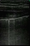
Resuscitative ultrasound image illustrating comet tail artifacts.
Figure 10
Figure 10
Resuscitative ultrasound image illustrating a lung point in time motion mode. The normal seashore sign (arrows) can be seen alternating with the stratosphere sign in time with respiration.
Figure 11
Figure 11
Resuscitative ultrasound image illustrating a subxiphoid view of the inferior vena cava (IVC) and hepatic vein (arrow). RA – right atrium, L – liver.
Figure 12
Figure 12
Resuscitative ultrasound image illustrating measurement of the optic nerve sheath diameter (3 mm behind the globe) in a patient with elevated intracranial pressure.

Similar articles

Cited by

References

    1. Research CIfH National trauma registry report: Hospital injury admissions 1998/1999. 2001.
    1. Krug EG, Sharma GK, Lozano R. The global burden of injuries. Am J Public Health. 2000;90:523–526. doi: 10.2105/AJPH.90.4.523. - DOI - PMC - PubMed
    1. Mathers CD, Loncar D. Projections of global mortality and burden of disease from 2002 to 2030. PLoS Med. 2006;3:e442. doi: 10.1371/journal.pmed.0030442. - DOI - PMC - PubMed
    1. Kirkpatrick AW, Sustic A, Blaivas M. Introduction to the use of ultrasound in critical care medicine. Crit Care Med. 2007;35:S123–S307. doi: 10.1097/01.CCM.0000260622.26564.94. - DOI
    1. Kirkpatrick AW, Ball CG, D'Amours SK, Zygun D. Acute resuscitation of the unstable adult trauma patient: bedside diagnosis and therapy. Can J Surg. 2008;51:57–69. - PMC - PubMed