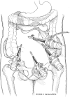
Laparoscopic repair of a right paraduodenal hernia
- PMID: 19660226
- PMCID: PMC3015939
Laparoscopic repair of a right paraduodenal hernia
Abstract
Background and objectives: Right paraduodenal hernia (PDH) results from a primitive gut malrotation. The resultant jejunal mesenteric defect posterior to the superior mesenteric vessels allows decompressed jejunum to herniate retroperitoneally. PDH make up 53% of all internal hernias, but account for only 0.2% to 5.8% of all cases of intestinal obstruction. In addition, PDH exhibits male and left-sided predominance. Ours is the second report to describe the preoperative diagnosis and totally laparoscopic repair of a right PDH.
Methods: We report the case of a 26-year-old female with symptoms suggestive of partial small bowel obstruction and a 6-year history of intermittent abdominal pain. Physical examination demonstrated lower quadrant tenderness. Plain abdominal radiographs and ultrasonography were nondiagnostic. Contrasted computed tomography of the abdomen revealed jejunum encased within the right upper quadrant suspicious for right PDH.
Results: The patient underwent successful laparoscopic right PDH repair and was discharged home on postoperative day 1 without late sequelae.
Conclusions: In the outpatient setting, clinical suspicion and comprehensive radiological investigation permit preoperative diagnosis of right PDH. In acute situations, clinical presentation, plain radiographs, and then diagnostic laparoscopy may be an expeditious diagnostic algorithm. Subsequent laparoscopic repair of right PDH is feasible and may shorten hospital length of stay.
Figures


References
-
- Blachar A, Federle MP. Internal hernia: an increasingly common cause of small bowel obstruction. Semin Ultrasound CT MR. 2002;23(2):174–183 - PubMed
-
- Berardi RS. Paraduodenal hernias. Surg Gynecol Obstet. 1981;152(1):99–110 - PubMed
-
- Yoo HY, Mergelas J, Seibert DG. Paraduodenal hernia: a treatable cause of upper gastrointestinal tract symptoms. J Clin Gastroenterol. 2000;31(3):226–229 - PubMed
-
- Brigham RA, Fallon WF, Saunders JR, Harmon JW, d'Avis JC. Paraduodenal hernia: diagnosis and surgical management. Surgery. 1984;96(3):498–502 - PubMed
-
- Khan MA, Lo AY, Van de Maele DM. Paraduodenal hernia. Am Surg. 1998;64(12):1218–1222 - PubMed
Publication types
MeSH terms
LinkOut - more resources
Full Text Sources
Medical
