

Minimally invasive spigelian hernia repair
- PMID: 19660230
- PMCID: PMC3015925
Minimally invasive spigelian hernia repair
Abstract
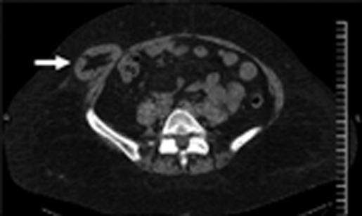
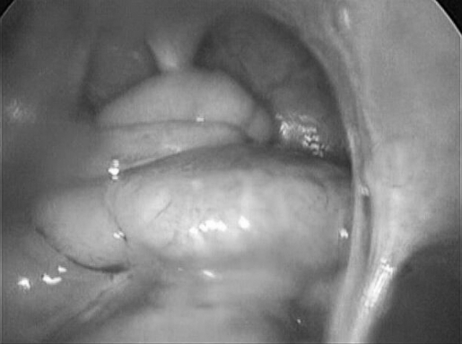
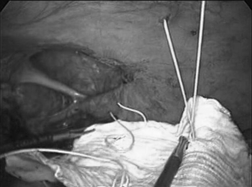
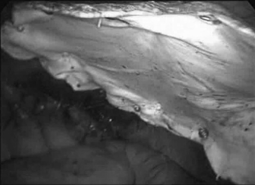
Introduction: Spigelian hernia is an uncommon ventral hernia characterized by a defect in the linea semilunaris. Repair of spigelian hernia has traditionally been accomplished via an open transverse incision and primary repair. The purpose of this article is to present 2 case reports of incarcerated spigelian hernia that were successfully repaired laparoscopically using Gortex mesh and to present a review of the literature regarding laparoscopic repair of spigelian hernias.
Methods: Retrospective chart review and Medline literature search.
Results: Two patients underwent laparoscopic mesh repair of incarcerated spigelian hernias. Both were started on a regular diet on postoperative day 1 and discharged on postoperative days 2 and 3. One patient developed a seroma that resolved without intervention. There was complete resolution of preoperative symptoms at the 12-month follow-up.
Conclusion: Minimally invasive repair of spigelian hernias is an alternative to the traditional open surgical technique. Further studies are needed to directly compare the open and the laparoscopic repair.
Figures
References
-
- Skandalakis PN, Zoras O, Skandalakis JE, Mirilas P. Spigelian hernia: surgical anatomy, embryology, and technique of repair. Am Surg. 2006;72:42–48 - PubMed
-
- Olson RO, Davis WC. Spigelian hernia: rare or obscure? Am J Surg. 1968;116:842–846 - PubMed
-
- Houlihan TJ. A review of spigelian hernia. Am J Surg. 1976;131:734–735 - PubMed
-
- Spangen L. Spigelian hernia. In: Prostheses and Abdominal Wall Hernias. Bendavid R. ed. Austin: RG Landes; 1994;563
-
- Spangen L. Spigelian hernia. World J Surg. 1989;13:573–580 - PubMed
Publication types
MeSH terms
LinkOut - more resources
Full Text Sources